 Explore
 Validate
 Learn
Learn Western blot
 ELISA
ELISA Immunoprecipitation
Antibody data
- Antibody Data
- Antigen structure
- References [0]
- Comments [0]
- Validations
- Western blot [2]
- Immunoprecipitation [3]
- Immunohistochemistry [6]
Submit
Validation data
Reference
Comment
Report error
- Product number
- LS-C377006 - Provider product page

- Provider
- LSBio
- Product name
- VTN / Vitronectin Antibody (aa364-478) LS-C377006
- Antibody type
- Polyclonal
- Description
- Immunoaffinity purified
- Reactivity
- Human
- Host
- Rabbit
- Isotype
- IgG
- Storage
- Short term: -20°C; Long term: -80°C; Avoid freeze-thaw cycles.
No comments: Submit comment
Enhanced validation
- Submitted by
- LSBio (provider)
- Enhanced method
- Genetic validation
- Main image
- Experimental details
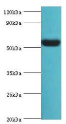
- Western blot. All lanes: Vitronectin antibody at 12 ug/ml+HepG2 whole cell lysate. Secondary antibody: Goat polyclonal to rabbit at 1:10000 dilution. Predicted band size: 54 kDa. Observed band size: 54 kDa Immunohistochemistry.
- Submitted by
- LSBio (provider)
- Enhanced method
- Genetic validation
- Main image

- Experimental details
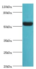
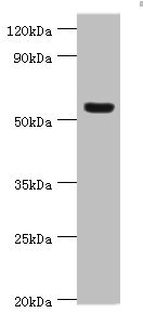
- Western blot All lanes: Vitronectin antibody at 12µg/ml + HepG2 whole cell lysate Secondary Goat polyclonal to rabbit IgG at 1/10000 dilution Predicted band size: 54 kDa Observed band size: 54 kDa
Supportive validation
- Submitted by
- LSBio (provider)
- Enhanced method
- Genetic validation
- Main image

- Experimental details
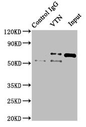
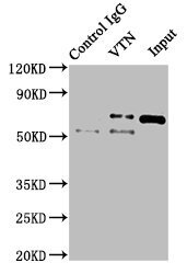
- Immunoprecipitating VTN in NIH/3T3 whole cell lysate Lane 1: Rabbit control IgG instead of VTN Antibody in NIH/3T3 whole cell lysate.For western blotting, a HRP-conjugated Protein G antibody was used as the secondary antibody (1/2000) Lane 2: VTN Antibody (8µg) + NIH/3T3 whole cell lysate (500µg) Lane 3: NIH/3T3 whole cell lysate (10µg)

- Submitted by
- LSBio (provider)
- Main image

- Experimental details
- Immunoprecipitating VTN in NIH/3T3 whole cell lysate Lane 1: Rabbit control IgG instead of VTN Antibody in NIH/3T3 whole cell lysate.For western blotting, a HRP-conjugated Protein G antibody was used as the secondary antibody (1/2000) Lane 2: VTN Antibody (8µg) + NIH/3T3 whole cell lysate (500µg) Lane 3: NIH/3T3 whole cell lysate (10µg)
- Submitted by
- LSBio (provider)
- Main image
- Experimental details
- Immunoprecipitating VTN in NIH/3T3 whole cell lysate Lane 1: Rabbit control IgG instead of VTN Antibody in NIH/3T3 whole cell lysate.For western blotting, a HRP-conjugated Protein G antibody was used as the secondary antibody (1/2000) Lane 2: VTN Antibody (8µg) + NIH/3T3 whole cell lysate (500µg) Lane 3: NIH/3T3 whole cell lysate (10µg)

Supportive validation
- Submitted by
- LSBio (provider)
- Enhanced method
- Genetic validation
- Main image

- Experimental details
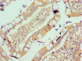
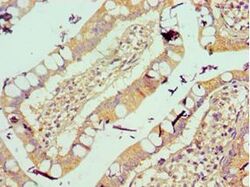
- Immunohistochemistry of paraffin-embedded human small intestine using antibody at 1:100 dilution.
- Submitted by
- LSBio (provider)
- Enhanced method
- Genetic validation
- Main image

- Experimental details
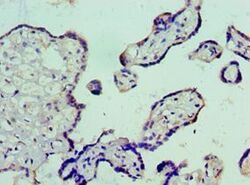
- Immunohistochemistry of paraffin-embedded human placenta using antibody at 1:100 dilution.

- Submitted by
- LSBio (provider)
- Enhanced method
- Genetic validation
- Main image

- Experimental details
- Immunohistochemistry of paraffin-embedded human small intestine using antibody at 1:100 dilution.

- Submitted by
- LSBio (provider)
- Enhanced method
- Genetic validation
- Main image
- Experimental details
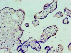
- Immunohistochemistry of paraffin-embedded human placenta using antibody at 1:100 dilution.
- Submitted by
- LSBio (provider)
- Enhanced method
- Genetic validation
- Main image
- Experimental details
- Immunohistochemistry of paraffin-embedded human small intestine using antibody at 1:100 dilution.

- Submitted by
- LSBio (provider)
- Enhanced method
- Genetic validation
- Main image

- Experimental details
- Immunohistochemistry of paraffin-embedded human placenta using antibody at 1:100 dilution.
