Explore
 Validate
 Learn
Learn Western blot
 ELISA
ELISA Immunohistochemistry
Antibody data
- Antibody Data
- Antigen structure
- References [2]
- Comments [0]
- Validations
- Immunohistochemistry [2]
- Other assay [4]
Submit
Validation data
Reference
Comment
Report error
- Product number
- PA5-27909 - Provider product page

- Provider
- Invitrogen Antibodies
- Product name
- Vitronectin Polyclonal Antibody
- Antibody type
- Polyclonal
- Antigen
- Recombinant full-length protein
- Description
- Recommended positive controls: HepG2 conditioned medium, mouse brain. Store product as a concentrated solution. Centrifuge briefly prior to opening the vial.
- Reactivity
- Human, Mouse
- Host
- Rabbit
- Isotype
- IgG
- Vial size
- 100 μL
- Concentration
- 0.34 mg/mL
- Storage
- Store at 4°C short term. For long term storage, store at -20°C, avoiding freeze/thaw cycles.
Submitted references Extracellular Matrix Proteins Confer Cell Adhesion-Mediated Drug Resistance Through Integrin α (v) in Glioblastoma Cells.
Candidate Glycoprotein Biomarkers for Canine Visceral Hemangiosarcoma and Validation Using Semi-Quantitative Lectin/Immunohistochemical Assays.
Yu Q, Xiao W, Sun S, Sohrabi A, Liang J, Seidlits SK
Frontiers in cell and developmental biology 2021;9:616580
Frontiers in cell and developmental biology 2021;9:616580
Candidate Glycoprotein Biomarkers for Canine Visceral Hemangiosarcoma and Validation Using Semi-Quantitative Lectin/Immunohistochemical Assays.
Oungsakul P, Choi E, Shah AK, Mohamed A, O'Leary C, Duffy D, Hill MM, Bielefeldt-Ohmann H
Veterinary sciences 2021 Feb 27;8(3)
Veterinary sciences 2021 Feb 27;8(3)
No comments: Submit comment
Supportive validation
- Submitted by
- Invitrogen Antibodies (provider)
- Main image
- Experimental details
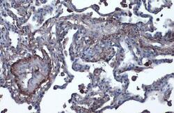
- Vitronectin Polyclonal Antibody detects Vitronectin protein by immunohistochemical analysis. Sample: Paraffin-embedded human lung cancer. Vitronectin stained by Vitronectin Polyclonal Antibody (Product # PA5-27909) diluted at 1:1,000. Antigen Retrieval: Citrate buffer, pH 6.0, 15 min.
- Submitted by
- Invitrogen Antibodies (provider)
- Main image

- Experimental details
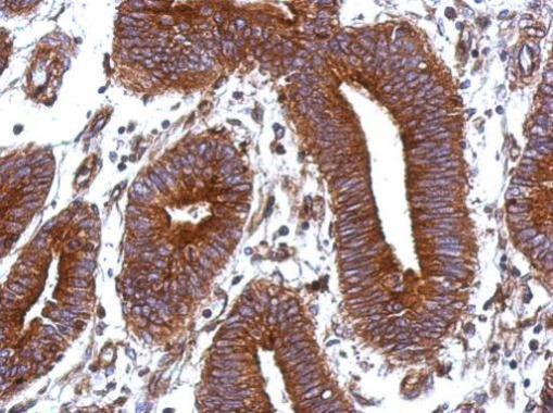
- Immunohistochemical analysis of paraffin-embedded human colon carcinoma, using Vitronectin (Product # PA5-27909) antibody at 1:500 dilution. Antigen Retrieval: EDTA based buffer, pH 8.0, 15 min.
Supportive validation
- Submitted by
- Invitrogen Antibodies (provider)
- Main image
- Experimental details
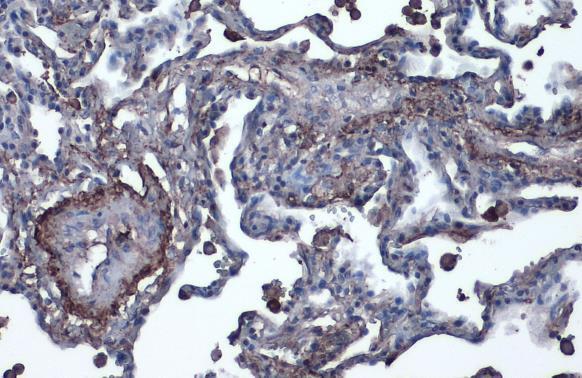
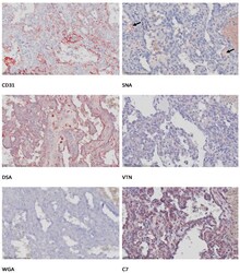
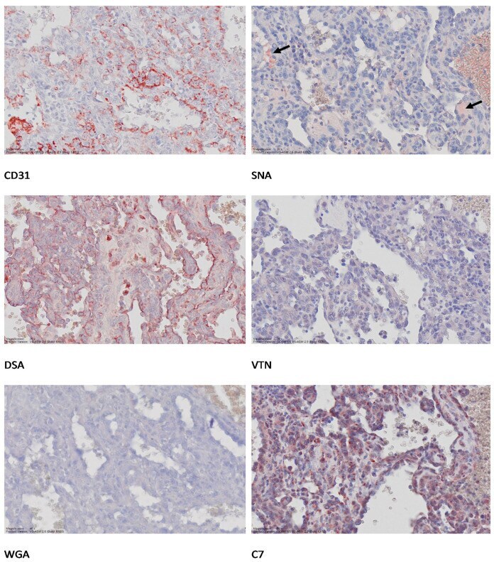
- Figure 2 Lectin-histochemistry and immunohistochemical labelling of canine splenic HSA. Formalin-fixed paraffin-embedded (FFPE) sections were reacted with anti-human CD31 antibody, anti-human VTN antibody, anti-human complement C7 antibody, and biotinylated lectins DSA ( Datura stramonium ), WGA (Wheat Germ Agglutinin), and SNA ( Sambucus nigra ) lectins) on consecutive splenic HSA sections (sample 13_019400E). Binding was visualized using AEC substrate (red) following either incubation with secondary antibody (Envision Kit) or streptavidin-horse radish peroxidase conjugate. Microphotos were generated using Aperio scans. Original magnifications 20x.
- Submitted by
- Invitrogen Antibodies (provider)
- Main image
- Experimental details
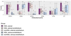
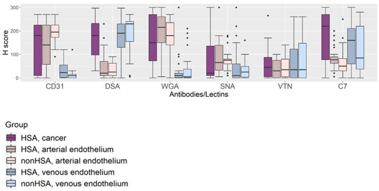
- Figure 3 Boxplot representing overall lectin-histochemistry (DSA, WGA, SNA) and immunohistochemical (CD31, VTN, C7) signal intensities. Three tissue components-HSA/cancer, arterial endothelium, and venous endothelium-were assessed in two sample groups-dogs with HSA and dogs without HSA. Differences in the level of tissue glycoprotein expression between tissue components and between HSA and non-HSA were compared.
- Submitted by
- Invitrogen Antibodies (provider)
- Main image
- Experimental details
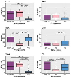
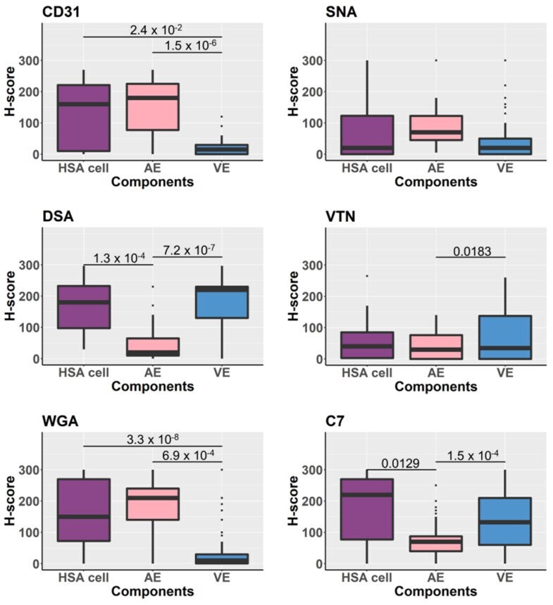
- Figure 4 Boxplots demonstrate the comparison of H-scores assessed from lectin-histochemistry (DSA, WGA, SNA) and immunohistochemical (CD31, VTN, C7) labelling of tissue samples. The statistical analysis was performed by comparing H-scores between tissue components-HSA/cancer cells, arterial endothelium (AE) and venous endothelium (VE). Significance bars show only p -values < 0.05 derived from Wilcoxon test and adjusted by Bonferroni corrections.
- Submitted by
- Invitrogen Antibodies (provider)
- Main image
- Experimental details
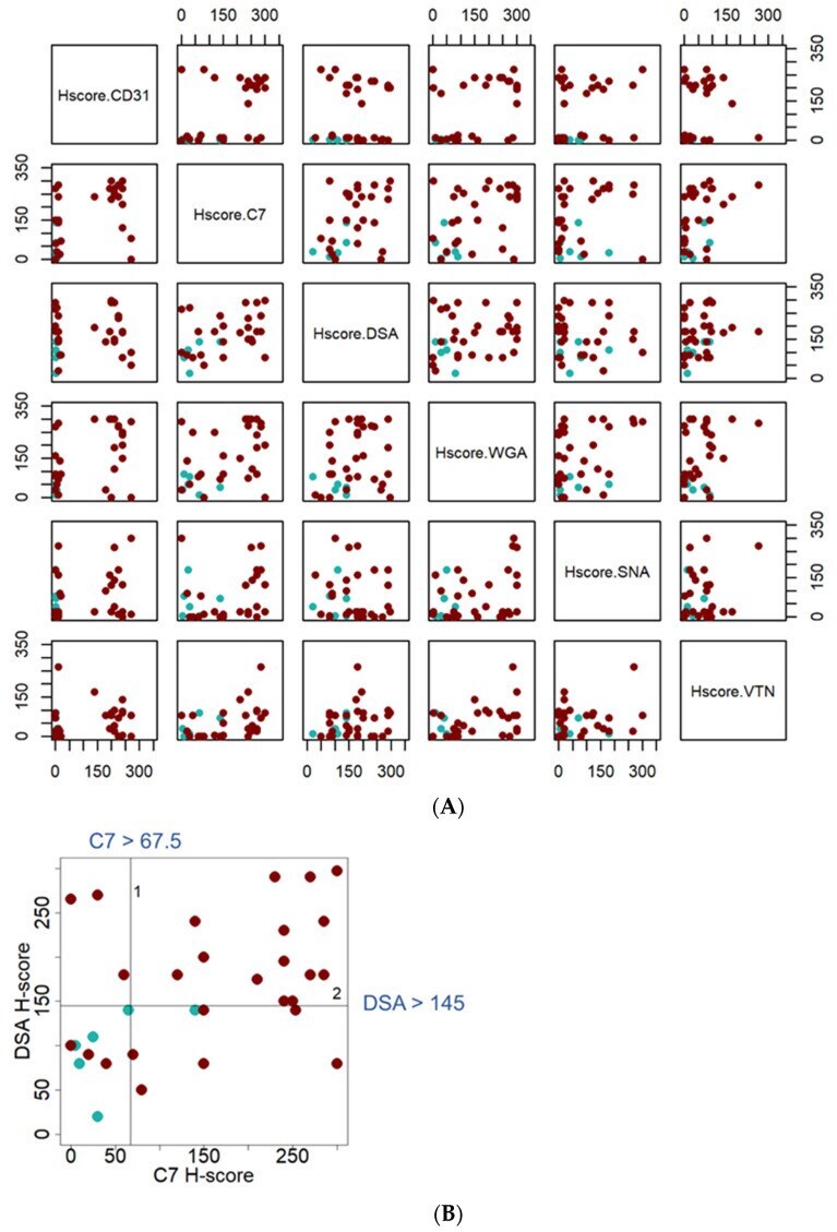
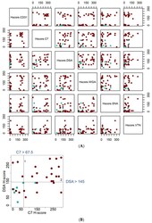
- Figure 5 Scatter plots of H-scores distribution, assessed from lectin-immunohistochemical labelling for complement C7 and VTN as well as DSA, WGA, and SNA. ( A ) Matrix scatter plots created for the candidate antibodies/lectins H-scores. The darker dots (dark brown) represent H-score assessed from the cancer component of HSA splenic tissue samples, while the lighter dots (light green) represent H-score assessed from the HSA-like lesion of non-HSA (hematoma and hemorrhage) splenic tissue samples. ( B ) Scatter plot of H-scores distribution assessed from lectin-immunohistochemical labelling with the anti-human complement C7 antibody and DSA lectin. The pair of reactants was selected from recursive partitioning results. Two cut-off points, complement C7 (H-score > 67.5) then DSA (H-score > 145), allow clear distinction between HSA and HSA-like tissues.