Explore
 Validate
 Learn
Learn Western blot
 Immunocytochemistry
Antibody data
- Antibody Data
- Antigen structure
- References [2]
- Comments [0]
- Validations
- Immunocytochemistry [1]
- Flow cytometry [1]
Submit
Validation data
Reference
Comment
Report error
- Product number
- 14-1029-37 - Provider product page

- Provider
- Invitrogen Antibodies
- Product name
- CD102 (ICAM-2) Monoclonal Antibody (CBR-IC2/2), eBioscience™
- Antibody type
- Monoclonal
- Antigen
- Other
- Description
- Description: The CBRIC2/2 monoclonal antibody reacts with human ICAM-2 (CD102). ICAM-2 is a 55-65 kD transmembrane glycoprotein possessing 2 extracellular Ig domains, a single transmembrane domain, and a short 26-amino acid cytoplasmic domain. ICAM-2 is expressed on most leukocytes, and is strongly expressed on vascular endothelial cells. ICAM-2 is a ligand of integrin CD11a/CD18 (LFA-1), and also has been reported to bind integrin CD11b/CD18 (Mac-1). ICAM-2 is an intercellular adhesion molecule involved in lymphocyte homing to sites of inflammation. The CBRIC2/2 antibody has been reported to have blocking activity. Applications Reported: This CBRIC2/2 antibody has been reported for use in flow cytometric analysis, immunoblotting, immunoprecipitation, immunohistochemical staining, and functional assays. Applications Tested: This CBRIC2/2 antibody has been tested by flow cytometric analysis of human peripheral blood cells. This can be used at less than or equal to 0.125 µg per test. A test is defined as the amount (µg) of antibody that will stain a cell sample in a final volume of 100 µL. Cell number should be determined empirically but can range from 10^5 to 10^8 cells/test. The CBRIC2/2 antibody has also been found useful for immunoblotting of a human peripheral blood cell lysate, recognizing a protein of approximately 55 kD under non-reducing conditions. It is recommended that the antibody be carefully titrated for optimal performance in the assay of interest. Purity: Greater than 90%, as determined by SDS-PAGE. Aggregation: Less than 10%, as determined by HPLC. Filtration: 0.2 µm post-manufacturing filtered.
- Reactivity
- Human
- Host
- Mouse
- Isotype
- IgG
- Antibody clone number
- CBR-IC2/2
- Vial size
- 2 mg
- Concentration
- 0.5 mg/mL
- Storage
- 4°C
Submitted references ICAM-1 and ICAM-2 Are Differentially Expressed and Up-Regulated on Inflamed Pulmonary Epithelium, but Neither ICAM-2 nor LFA-1: ICAM-1 Are Required for Neutrophil Migration Into the Airways In Vivo.
Identification of endothelial cell junctional proteins and lymphocyte receptors involved in transendothelial migration of human effector memory CD4+ T cells.
Chong DLW, Rebeyrol C, José RJ, Williams AE, Brown JS, Scotton CJ, Porter JC
Frontiers in immunology 2021;12:691957
Frontiers in immunology 2021;12:691957
Identification of endothelial cell junctional proteins and lymphocyte receptors involved in transendothelial migration of human effector memory CD4+ T cells.
Manes TD, Pober JS
Journal of immunology (Baltimore, Md. : 1950) 2011 Feb 1;186(3):1763-8
Journal of immunology (Baltimore, Md. : 1950) 2011 Feb 1;186(3):1763-8
No comments: Submit comment
Supportive validation
- Submitted by
- Invitrogen Antibodies (provider)
- Main image
- Experimental details
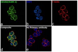
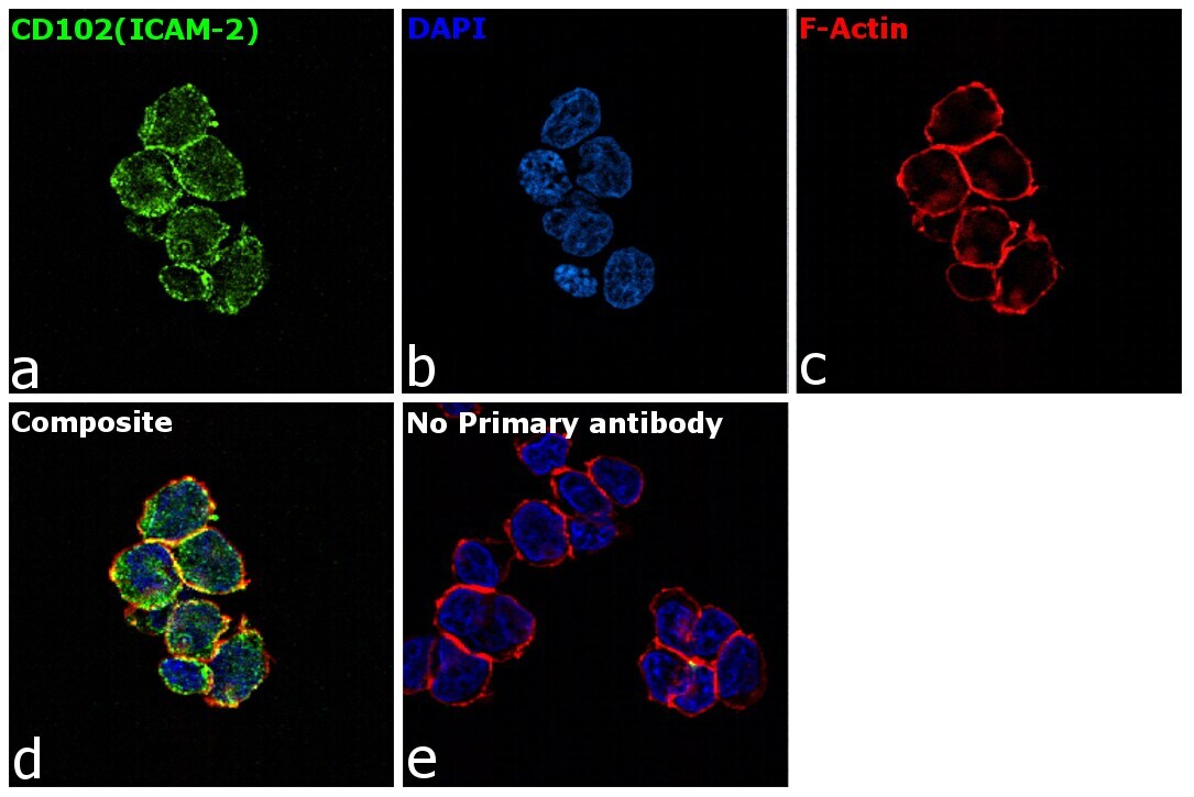
- Immunofluorescence analysis of CD102 (ICAM-2) was performed using log phase Jurkat cells. The cells were fixed with 4% paraformaldehyde for 10 minutes, permeabilized with 0.1% Triton™ X-100 for 15 minutes, and blocked with 2% BSA for 1 hour at room temperature. The cells were labeled with CD102 (ICAM-2) Mouse Monoclonal Antibody (CBR-IC2/2) (Product # 14-1029-82) at 5 µg/mL in 0.1% BSA, incubated at 4 degree Celsius overnight and then with Goat anti-Mouse IgG (H+L) Superclonal™ Recombinant Secondary Antibody, Alexa Fluor® 488 conjugate (Product # A28175) at a dilution of 1:2000 for 45 minutes at room temperature (Panel a: green). Nuclei (Panel b: blue) were stained with ProLong™ Diamond Antifade Mountant with DAPI (Product # P36962). F-actin (Panel c: red) was stained with Rhodamine Phalloidin (Product # R415, 1:300). Panel d represents the merged image showing membrane localization. Panel e represents control cells with no primary antibody to assess background. The images were captured at 60X magnification.
Supportive validation
- Submitted by
- Invitrogen Antibodies (provider)
- Main image
- Experimental details
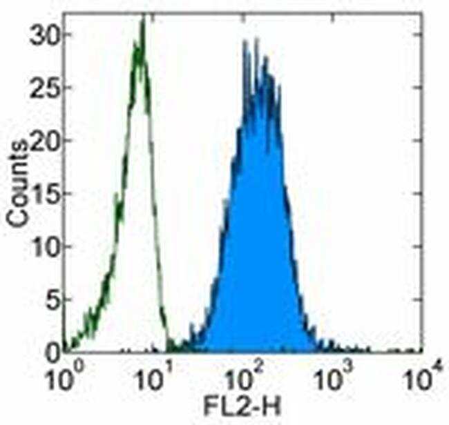
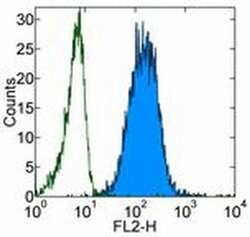
- Staining of normal human peripheral blood cells with 0.06 µg of Mouse IgG2a kappa Isotype Control Purified (Product # 14-4724-82) (open histogram) or 0.06 µg of Anti-Human CD102 (ICAM-2) Purified (filled histogram) followed by F (ab')2 Anti-Mouse IgG PE (Product # 12-4012). Cells in the monocyte gate were used for analysis.