 Explore
 Validate
 Learn
Learn Western blot
 Immunocytochemistry
 Immunohistochemistry
Antibody data
- Antibody Data
- Antigen structure
- References [3]
- Comments [0]
- Validations
- Immunocytochemistry [4]
- Immunoprecipitation [1]
- Other assay [3]
Submit
Validation data
Reference
Comment
Report error
- Product number
- MA1-26736 - Provider product page

- Provider
- Invitrogen Antibodies
- Product name
- hnRNP A1 Monoclonal Antibody (9H10)
- Antibody type
- Monoclonal
- Antigen
- Purifed from natural sources
- Description
- Recommended positive controls: 293T. A band of approximately 34 kDa is detected by Western Blot.
- Reactivity
- Human, Mouse, Rat, Bovine, Canine, Hamster
- Host
- Mouse
- Isotype
- IgG
- Antibody clone number
- 9H10
- Vial size
- 50 μL
- Concentration
- 2.2 mg/mL
- Storage
- Store at 4°C short term. For long term storage, store at -20°C, avoiding freeze/thaw cycles.
Submitted references Comparative analyses of alphaviral RNA:Protein complexes reveals conserved host-pathogen interactions.
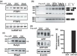
MicroRNA-132 loss is associated with tau exon 10 inclusion in progressive supranuclear palsy.
In vivo regulation of amyloid precursor protein neuronal splicing by microRNAs.
Gebhart NN, Hardy RW, Sokoloski KJ
PloS one 2020;15(8):e0238254
PloS one 2020;15(8):e0238254
MicroRNA-132 loss is associated with tau exon 10 inclusion in progressive supranuclear palsy.
Smith PY, Delay C, Girard J, Papon MA, Planel E, Sergeant N, Buée L, Hébert SS
Human molecular genetics 2011 Oct 15;20(20):4016-24
Human molecular genetics 2011 Oct 15;20(20):4016-24
In vivo regulation of amyloid precursor protein neuronal splicing by microRNAs.
Smith P, Al Hashimi A, Girard J, Delay C, Hébert SS
Journal of neurochemistry 2011 Jan;116(2):240-7
Journal of neurochemistry 2011 Jan;116(2):240-7
No comments: Submit comment
Supportive validation
- Submitted by
- Invitrogen Antibodies (provider)
- Main image

- Experimental details
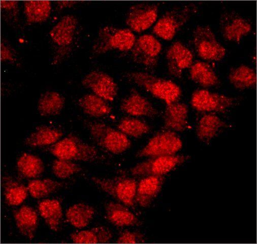
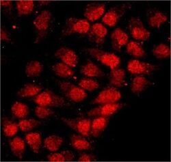
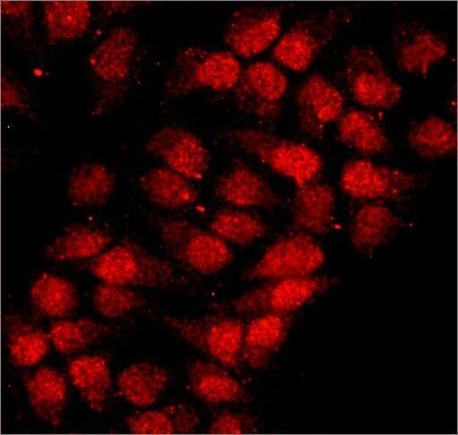
- Immunocytochemistry-Immunofluorescence analysis of hnRNP A1 in HeLa cells using hnRNP A1 Monoclonal Antibody (9H10) (Product # MA1 26736) at 10 µg/mL. Cells were fixed and permeabilized with cold methanol followed by cold methanol: acetone.
- Submitted by
- Invitrogen Antibodies (provider)
- Main image

- Experimental details
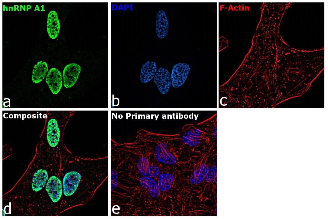
- Immunofluorescence analysis of hnRNP A1 was performed using 70% confluent log phase HeLa cells. The cells were fixed with 4% paraformaldehyde for 10 minutes, permeabilized with 0.1% Triton™ X-100 for 15 minutes, and blocked with 1% BSA for 1 hour at room temperature. The cells were labeled with hnRNP A1 Monoclonal Antibody (9H10) (Product # MA1-26736) at 5 microgram/mL in 0.1% BSA, incubated at 4 degree Celsius overnight and then labeled with Goat anti-Mouse IgG (H+L) Superclonal™ Secondary Antibody, Alexa Fluor® 488 conjugate (Product # A28175) at a dilution of 1:2000 for 45 minutes at room temperature (Panel a: green). Nuclei (Panel b: blue) were stained with ProLong™ Diamond Antifade Mountant with DAPI (Product # P36962). F-actin (Panel c: red) was stained with Rhodamine Phalloidin (Product # R415, 1:300). Panel d represents the merged image showing Nuclear localization. Panel e represents control cells with no primary antibody to assess background. The images were captured at 60X magnification.
- Submitted by
- Invitrogen Antibodies (provider)
- Main image

- Experimental details
- Immunofluorescence analysis of hnRNP A1 was performed using 70% confluent log phase HeLa cells. The cells were fixed with 4% paraformaldehyde for 10 minutes, permeabilized with 0.1% Triton™ X-100 for 15 minutes, and blocked with 1% BSA for 1 hour at room temperature. The cells were labeled with hnRNP A1 Monoclonal Antibody (9H10) (Product # MA1-26736) at 5 microgram/mL in 0.1% BSA, incubated at 4 degree Celsius overnight and then labeled with Goat anti-Mouse IgG (H+L) Superclonal™ Secondary Antibody, Alexa Fluor® 488 conjugate (Product # A28175) at a dilution of 1:2000 for 45 minutes at room temperature (Panel a: green). Nuclei (Panel b: blue) were stained with ProLong™ Diamond Antifade Mountant with DAPI (Product # P36962). F-actin (Panel c: red) was stained with Rhodamine Phalloidin (Product # R415, 1:300). Panel d represents the merged image showing Nuclear localization. Panel e represents control cells with no primary antibody to assess background. The images were captured at 60X magnification.

- Submitted by
- Invitrogen Antibodies (provider)
- Main image
- Experimental details
- Immunocytochemistry-Immunofluorescence analysis of hnRNP A1 in HeLa cells using hnRNP A1 Monoclonal Antibody (9H10) (Product # MA1 26736) at 10 µg/mL. Cells were fixed and permeabilized with cold methanol followed by cold methanol: acetone.
Supportive validation
- Submitted by
- Invitrogen Antibodies (provider)
- Main image

- Experimental details
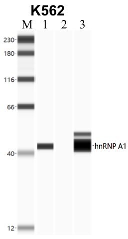
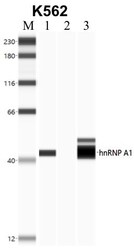
- Immunoprecipitation of hnRNP A1 was performed in K562 cells. Antigen-antibody complexes were formed by incubating approximately 500 µg whole cell lysate with 5 to 10 µL of monoclonal hnRNP A1 antibody (Product # MA1-26736) rotating 60 min at RT. The immune complexes were captured on 625 µg of anti-mouse coated Dynabeads (Product # 11202D) and washed extensively. They were then eluted and analyzed using the Simple Western system using the same antibody as used in immunoprecipitation at a dilution of 1:25, followed by a 1:100 dilution of secondary antibody. Lane 1 is the input, lane 2 no antibody IP and lane 3 is the target specific IP. Data courtesy of the Yeo lab as part of the ENCODE project.
Supportive validation
- Submitted by
- Invitrogen Antibodies (provider)
- Main image
- Experimental details
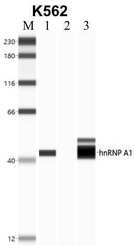
- RNA immunoprecipitation (RIP) western of hnRNP A1 was performed in K562 cells. Antigen-antibody complexes were formed by incubating approximately 500 µg whole cell lysate with 5 to 10 µL of monoclonal hnRNP A1 antibody (Product # MA1-26736) rotating 60 min at RT. The immune complexes were captured on 625 µg of anti-mouse coated Dynabeads (Product # 11202D) and washed extensively. They were then eluted and analyzed using the Simple Western system using the same antibody as used in immunoprecipitation at a dilution of 1:25, followed by a 1:100 dilution of secondary antibody. Lane 1 is the input, lane 2 no antibody IP and lane 3 is the target specific IP. Data courtesy of the Yeo lab as part of the ENCODE project.
- Submitted by
- Invitrogen Antibodies (provider)
- Main image
- Experimental details
- NULL

- Submitted by
- Invitrogen Antibodies (provider)
- Main image
- Experimental details
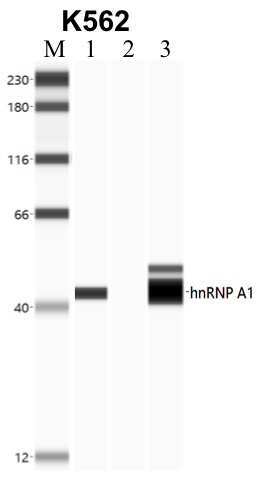
- RNA immunoprecipitation (RIP) western of hnRNP A1 was performed in K562 cells. Antigen-antibody complexes were formed by incubating approximately 500 µg whole cell lysate with 5 to 10 µL of monoclonal hnRNP A1 antibody (Product # MA1-26736) rotating 60 min at RT. The immune complexes were captured on 625 µg of anti-mouse coated Dynabeads (Product # 11202D) and washed extensively. They were then eluted and analyzed using the Simple Western system using the same antibody as used in immunoprecipitation at a dilution of 1:25, followed by a 1:100 dilution of secondary antibody. Lane 1 is the input, lane 2 no antibody IP and lane 3 is the target specific IP. Data courtesy of the Yeo lab as part of the ENCODE project.
