 Explore
 Validate
 Learn
Learn Western blot
 ELISA
ELISAAntibody data
- Antibody Data
- Antigen structure
- References [0]
- Comments [0]
- Validations
- Western blot [4]
- Immunocytochemistry [2]
Submit
Validation data
Reference
Comment
Report error
- Product number
- MA5-24774 - Provider product page

- Provider
- Invitrogen Antibodies
- Product name
- hnRNP A1 Monoclonal Antibody (4B10)
- Antibody type
- Monoclonal
- Antigen
- Purifed from natural sources
- Description
- Does not react with hnRNP A2 or B.
- Reactivity
- Human, Mouse, Bovine, Canine
- Host
- Mouse
- Isotype
- IgG
- Antibody clone number
- 4B10
- Vial size
- 100 µL
- Concentration
- 1 mg/mL
- Storage
- Store at 4°C short term. For long term storage, store at -20°C, avoiding freeze/thaw cycles.
No comments: Submit comment
Supportive validation
- Submitted by
- Invitrogen Antibodies (provider)
- Main image
- Experimental details
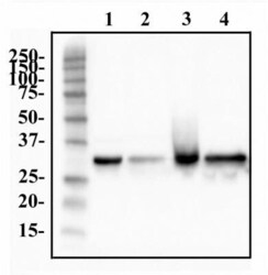
- Western blot analysis of hnRNP A1 in whole cell protein from HeLa, A431, Hek293, and MCF7. Samples were incubated in hnRNP A1 monoclonal antibody (Product # MA5-24774) using a dilution of 0.5 mg/mL followed by an anti-mouse HRP secondary antibody secondary antibody. Separated on a 12% gel by SDS-PAGE, transferred to PVDF membrane and blocked in 5% non-fat milk in TBST. Precision Plus Protein All Blue molecular weight markers were detected with 1 µg/mL Anti-Blue Marker antibody. Detection: chemiluminescence.

- Submitted by
- Invitrogen Antibodies (provider)
- Main image

- Experimental details
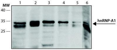
- Western blot analysis of hnRNP A1 in Lane 1: HeLa; Lane 2: Jurkat; Lane 3: HEK-293T, Lane 4: WiDr, Lane 5: NCI-H1299, Lane 6: F9. Samples were incubated in hnRNP A1 monoclonal antibody (Product # MA5-24774).
- Submitted by
- Invitrogen Antibodies (provider)
- Main image
- Experimental details
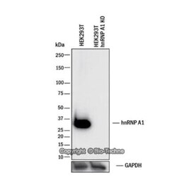
- Knockout validation by Western blot analysis of hnRNP A1 in lysates of HEK293T human embryonic kidney parental cell line and hnRNP A1 knockout (KO) HEK293T cell line. Samples were incubated in hnRNP A1 monoclonal antibody (Product # MA5-24774) using a dilution of 0.5 µg/mL followed by a HRP-conjugated Anti-Mouse IgG secondary antibody. Specific band was detected for hnRNP A1 at approximately 35 kDa (as indicated) in the parental HEK293T cell line, but is not detectable in the knockout HEK293T cell line. This experiment was conducted under reducing conditions.

- Submitted by
- Invitrogen Antibodies (provider)
- Main image
- Experimental details
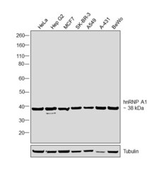
- Western blot was performed using Anti-hnRNP A1 Monoclonal Antibody (4B10), (Product # MA5-24774) and a 38 kDa band corresponding to hnRNP A1 was observed in the cell lines tested. Modified whole cell extracts (1%SDS) (30 µg lysate) of HeLa (Lane 1), Hep G2 (Lane 2), MCF7 (Lane 3), SK-BR-3 (Lane 4), A549 (Lane 5), A-431 (Lane 6) and BeWo (Lane 7) were electrophoresed using Novex® NuPAGE® 4-12 % Bis-Tris gel (Product # NP0321BOX). Resolved proteins were then transferred onto a nitrocellulose membrane (Product # IB23001) by iBlot® 2 Dry Blotting System (Product # IB21001). The blot was probed with the primary antibody (1 µg/mL) and detected by chemiluminescence with Goat anti-Mouse IgG (H+L), Superclonal™ Recombinant Secondary Antibody, HRP (Product # A28177, 1:4000 dilution) using the iBright FL 1000 (Product # A32752). Chemiluminescent detection was performed using Novex® ECL Chemiluminescent Substrate Reagent Kit (Product # WP20005).

Supportive validation
- Submitted by
- Invitrogen Antibodies (provider)
- Main image

- Experimental details
- Immunofluorescent analysis of hnRNP A1 in HeLa cells using hnRNP A1 monoclonal antibody (Product # MA5-24774) with a dilution of 1:200, Phalloidin 568 (red) and DAPI (blue).

- Submitted by
- Invitrogen Antibodies (provider)
- Main image

- Experimental details
- Immunofluorescence analysis of HNRNPA1 was performed using 70% confluent log phase MCF7 cells. The cells were fixed with 4% paraformaldehyde for 10 minutes, permeabilized with 0.1% Triton™ X-100 for 15 minutes, and blocked with 2% BSA for 1 hour at room temperature. The cells were labeled with hnRNP A1 Monoclonal Antibody (4B10) (Product # MA5-24774) at 1:200 dilution in 0.1% BSA, incubated at 4 degree celsius overnight and then with Donkey anti-Mouse IgG (H+L) Highly Cross-Adsorbed Secondary Antibody, Alexa Fluor Plus 488 (Product # A32766) at a dilution of 1:2000 for 45 minutes at room temperature (Panel a: green). Nuclei (Panel b: blue) were stained with SlowFade® Gold Antifade Mountant with DAPI (Product # S36938). F-actin (Panel c: red) was stained with Rhodamine Phalloidin (Product # R415, 1:300). Panel d represents the merged image showing staining in nucleus. Panel e represents control cells with no primary antibody to assess background. The images were captured at 60X magnification.
