 Explore
 Validate
 Learn
Learn Flow cytometry
Antibody data
- Antibody Data
- Antigen structure
- References [14]
- Comments [0]
- Validations
- Flow cytometry [1]
- Other assay [8]
Submit
Validation data
Reference
Comment
Report error
- Product number
- MHCD1406 - Provider product page

- Provider
- Invitrogen Antibodies
- Product name
- CD14 Monoclonal Antibody (TuK4), TRI-COLOR™
- Antibody type
- Monoclonal
- Antigen
- Other
- Description
- The TRI-COLOR® conjugate is a tandem R-phycoerythrin (PE)-Cy5® conjugate that permits simultaneous multicolor labeling and detection of multiple targets with excitation by a single excitation source-the 488 nm line of the argon-ion laser.
- Reactivity
- Human
- Host
- Mouse
- Isotype
- IgG
- Antibody clone number
- TuK4
- Vial size
- 500 µL
- Storage
- 4° C, store in dark
Submitted references Adjuvanted HIV-1 vaccine promotes antibody-dependent phagocytic responses and protects against heterologous SHIV challenge.
A molecular cell atlas of the human lung from single-cell RNA sequencing.
Antibodies to a Conserved Influenza Head Interface Epitope Protect by an IgG Subtype-Dependent Mechanism.
Ultra-High-Frequency Reprogramming of Individual Long-Term Hematopoietic Stem Cells Yields Low Somatic Variant Induced Pluripotent Stem Cells.
CBX7 Induces Self-Renewal of Human Normal and Malignant Hematopoietic Stem and Progenitor Cells by Canonical and Non-canonical Interactions.
IL1RAP potentiates multiple oncogenic signaling pathways in AML.
Eradication of Canine Diffuse Large B-Cell Lymphoma in a Murine Xenograft Model with CD47 Blockade and Anti-CD20.
Eltrombopag inhibits the proliferation of leukemia cells via reduction of intracellular iron and induction of differentiation.
Cytokine signatures of innate and adaptive immunity in 17DD yellow fever vaccinated children and its association with the level of neutralizing antibody.
Uncoupling of proliferation and cytokines from suppression within the CD4+CD25+Foxp3+ T-cell compartment in the 1st year of human type 1 diabetes.
Abnormal cytokine production by bone marrow stromal cells of multiple myeloma patients in response to RPMI8226 myeloma cells.
Blister fluid T lymphocytes during toxic epidermal necrolysis are functional cytotoxic cells which express human natural killer (NK) inhibitory receptors.
Blister fluid T lymphocytes during toxic epidermal necrolysis are functional cytotoxic cells which express human natural killer (NK) inhibitory receptors.
The first alpha helix of interleukin (IL)-2 folds as a homotetramer, acts as an agonist of the IL-2 receptor beta chain, and induces lymphokine-activated killer cells.
Om K, Paquin-Proulx D, Montero M, Peachman K, Shen X, Wieczorek L, Beck Z, Weiner JA, Kim D, Li Y, Mdluli T, Shubin Z, Bryant C, Sharma V, Tokarev A, Dawson P, White Y, Appelbe O, Klatt NR, Tovanabutra S, Estes JD, Matyas GR, Ferrari G, Alving CR, Tomaras GD, Ackerman ME, Michael NL, Robb ML, Polonis V, Rolland M, Eller MA, Rao M, Bolton DL
PLoS pathogens 2020 Sep;16(9):e1008764
PLoS pathogens 2020 Sep;16(9):e1008764
A molecular cell atlas of the human lung from single-cell RNA sequencing.
Travaglini KJ, Nabhan AN, Penland L, Sinha R, Gillich A, Sit RV, Chang S, Conley SD, Mori Y, Seita J, Berry GJ, Shrager JB, Metzger RJ, Kuo CS, Neff N, Weissman IL, Quake SR, Krasnow MA
Nature 2020 Nov;587(7835):619-625
Nature 2020 Nov;587(7835):619-625
Antibodies to a Conserved Influenza Head Interface Epitope Protect by an IgG Subtype-Dependent Mechanism.
Watanabe A, McCarthy KR, Kuraoka M, Schmidt AG, Adachi Y, Onodera T, Tonouchi K, Caradonna TM, Bajic G, Song S, McGee CE, Sempowski GD, Feng F, Urick P, Kepler TB, Takahashi Y, Harrison SC, Kelsoe G
Cell 2019 May 16;177(5):1124-1135.e16
Cell 2019 May 16;177(5):1124-1135.e16
Ultra-High-Frequency Reprogramming of Individual Long-Term Hematopoietic Stem Cells Yields Low Somatic Variant Induced Pluripotent Stem Cells.
Wang K, Guzman AK, Yan Z, Zhang S, Hu MY, Hamaneh MB, Yu YK, Tolu S, Zhang J, Kanavy HE, Ye K, Bartholdy B, Bouhassira EE
Cell reports 2019 Mar 5;26(10):2580-2592.e7
Cell reports 2019 Mar 5;26(10):2580-2592.e7
CBX7 Induces Self-Renewal of Human Normal and Malignant Hematopoietic Stem and Progenitor Cells by Canonical and Non-canonical Interactions.
Jung J, Buisman SC, Weersing E, Dethmers-Ausema A, Zwart E, Schepers H, Dekker MR, Lazare SS, Hammerl F, Skokova Y, Kooistra SM, Klauke K, Poot RA, Bystrykh LV, de Haan G
Cell reports 2019 Feb 12;26(7):1906-1918.e8
Cell reports 2019 Feb 12;26(7):1906-1918.e8
IL1RAP potentiates multiple oncogenic signaling pathways in AML.
Mitchell K, Barreyro L, Todorova TI, Taylor SJ, Antony-Debré I, Narayanagari SR, Carvajal LA, Leite J, Piperdi Z, Pendurti G, Mantzaris I, Paietta E, Verma A, Gritsman K, Steidl U
The Journal of experimental medicine 2018 Jun 4;215(6):1709-1727
The Journal of experimental medicine 2018 Jun 4;215(6):1709-1727
Eradication of Canine Diffuse Large B-Cell Lymphoma in a Murine Xenograft Model with CD47 Blockade and Anti-CD20.
Weiskopf K, Anderson KL, Ito D, Schnorr PJ, Tomiyasu H, Ring AM, Bloink K, Efe J, Rue S, Lowery D, Barkal A, Prohaska S, McKenna KM, Cornax I, O'Brien TD, O'Sullivan MG, Weissman IL, Modiano JF
Cancer immunology research 2016 Dec;4(12):1072-1087
Cancer immunology research 2016 Dec;4(12):1072-1087
Eltrombopag inhibits the proliferation of leukemia cells via reduction of intracellular iron and induction of differentiation.
Roth M, Will B, Simkin G, Narayanagari S, Barreyro L, Bartholdy B, Tamari R, Mitsiades CS, Verma A, Steidl U
Blood 2012 Jul 12;120(2):386-94
Blood 2012 Jul 12;120(2):386-94
Cytokine signatures of innate and adaptive immunity in 17DD yellow fever vaccinated children and its association with the level of neutralizing antibody.
Luiza-Silva M, Campi-Azevedo AC, Batista MA, Martins MA, Avelar RS, da Silveira Lemos D, Bastos Camacho LA, de Menezes Martins R, de Lourdes de Sousa Maia M, Guedes Farias RH, da Silva Freire M, Galler R, Homma A, Leite Ribeiro JG, Campos Lemos JA, Auxiliadora-Martins M, Eloi-Santos SM, Teixeira-Carvalho A, Martins-Filho OA
The Journal of infectious diseases 2011 Sep 15;204(6):873-83
The Journal of infectious diseases 2011 Sep 15;204(6):873-83
Uncoupling of proliferation and cytokines from suppression within the CD4+CD25+Foxp3+ T-cell compartment in the 1st year of human type 1 diabetes.
Hughson A, Bromberg I, Johnson B, Quataert S, Jospe N, Fowell DJ
Diabetes 2011 Aug;60(8):2125-33
Diabetes 2011 Aug;60(8):2125-33
Abnormal cytokine production by bone marrow stromal cells of multiple myeloma patients in response to RPMI8226 myeloma cells.
Zdzisińska B, Bojarska-Junak A, Dmoszyńska A, Kandefer-Szerszeń M
Archivum immunologiae et therapiae experimentalis 2008 May-Jun;56(3):207-21
Archivum immunologiae et therapiae experimentalis 2008 May-Jun;56(3):207-21
Blister fluid T lymphocytes during toxic epidermal necrolysis are functional cytotoxic cells which express human natural killer (NK) inhibitory receptors.
Le Cleach L, Delaire S, Boumsell L, Bagot M, Bourgault-Villada I, Bensussan A, Roujeau JC
Clinical and experimental immunology 2000 Jan;119(1):225-30
Clinical and experimental immunology 2000 Jan;119(1):225-30
The first alpha helix of interleukin (IL)-2 folds as a homotetramer, acts as an agonist of the IL-2 receptor beta chain, and induces lymphokine-activated killer cells.
Eckenberg R, Rose T, Moreau JL, Weil R, Gesbert F, Dubois S, Tello D, Bossus M, Gras H, Tartar A, Bertoglio J, Chouaïb S, Goldberg M, Jacques Y, Alzari PM, Thèze J
The Journal of experimental medicine 2000 Feb 7;191(3):529-40
The Journal of experimental medicine 2000 Feb 7;191(3):529-40
No comments: Submit comment
Supportive validation
- Submitted by
- Invitrogen Antibodies (provider)
- Main image

- Experimental details
- Human peripheral blood monocytes stained using TRI-COLOR® of anti-human CD14 monoclonal antibody (clone Tuk4). The negative control profiles represent unstained cells.

Supportive validation
- Submitted by
- Invitrogen Antibodies (provider)
- Main image

- Experimental details
- NULL

- Submitted by
- Invitrogen Antibodies (provider)
- Main image

- Experimental details
- NULL

- Submitted by
- Invitrogen Antibodies (provider)
- Main image
- Experimental details
- NULL

- Submitted by
- Invitrogen Antibodies (provider)
- Main image

- Experimental details
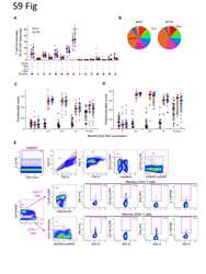
- Figure 1. Targeting of IL1RAP reduces growth of human AML cells by inducing differentiation and apoptosis, without affecting healthy hematopoietic cells. (A) Cell proliferation of THP-1 AML cells with replenishment of IL1RAP polyclonal antibody (pAb). 100 ug/ml of each antibody was added at day 0 and where indicated by the symbol +. Data represent the mean +- SD of two independent experiments. P-values were calculated using unpaired two-tailed t tests, and multiple comparisons were corrected for using the Holm-Sidak method. (B) Cell proliferation of THP-1 cells treated with different doses of IL1RAP pAb. Data represent the mean +- SD of three independent experiments. P-values were calculated using unpaired two-tailed t tests and multiple comparisons were corrected for using the Holm-Sidak method. (C) Morphology of THP-1 cells after treatment with 150 ug/ml IL1RAP pAb or isotype control pAb for 24 h. (D) Expression of macrophage differentiation markers in THP-1 cells by flow cytometry 24 h after addition of 150 ug/ml IL1RAP pAb. Representative histograms are shown (top). Bar graphs (bottom) represent the mean +- SD of five independent experiments. P-values were calculated using ratio paired two-tailed t tests comparing raw MFI values of isotype control pAb- versus IL1RAP pAb-treated cells. (E) Percentage of apoptotic (annexinV + ) THP-1 cells after treatment with 150 ug/ml IL1RAP pAb for 72 h. Data represent the mean +- SD of five independent experiments. P-values were calcula

- Submitted by
- Invitrogen Antibodies (provider)
- Main image

- Experimental details
- 10.1371/journal.ppat.1008764.g002 Fig 2 Env-specific antibody binding and effector responses elicited by vaccination. (A) Serum IgG binding antibody responses to gp145 and SHIV-1157ipd3N4 antigens are shown at peak and time of challenge. Area under the curve (AUC) reflects BAMA MFI values across serially diluted serum. (B) Peak rectal binding IgG responses reported as specific activity (SA), calculated as MFI * dilution / IgG concentration. All active arm responses in (A-B) are significantly different from the control arm, but not from each other. (C) Longitudinal serum ADCC responses were assessed by antibody-dependent lysis of CFSE-labeled Env-coated target cells and are reported as an increase in CFSE-negative target cells. (D) Antibody-dependent NK cell responses were measured by Env stimulation in the presence of animal sera followed by intracellular cytokine staining (ICS) for IFNgamma, TNFalpha, and MIP1beta. The frequency of NK cells expressing one or more cytokine Boolean combinations is shown. (E) ADCP and ADNP responses were assessed using THP-1 monocyte and human neutrophil effectors, respectively. ADNP open symbol depicts animal that resisted three additional high-dose challenges. Effector assays were performed with CO6980v0c22 multimeric gp145. For ADCP and ADNP, serum was used at month 0 and plasma at months 12.7 and 15 due to sample availability. Gray bars reflect interquartile range; black lines depict medians; P values in (C-E) reflect Wilcoxon rank-sum test

- Submitted by
- Invitrogen Antibodies (provider)
- Main image

- Experimental details
- 10.1371/journal.ppat.1008764.g003 Fig 3 HIV-specific CD4 and CD8 T cell responses. (A) PBMC HIV-1 Env-specific CD4 and CD8 T cells were measured by ICS for IFNgamma, TNFalpha, IL-2, and CD154 three weeks following the third (month 6.7) and fourth (month 12.7) immunizations. The frequency of memory cells that expressed one or more cytokines following ex vivo Env PTE peptide pool stimulation is shown for each animal by arm. CD4 responses represent summation of responses to pools 1-3 (spanning amino acids 4-585), while CD8 are to pool 3 only (amino acids 413-585). (B) Bronchoalveolar lavage Env-specific CD4 and CD8 T cells were measured by ICS four weeks post-infection for animals infected by challenges one through ten. Gray bars depict interquartile range; black lines depict medians; P values reflect Wilcoxon rank-sum test.

- Submitted by
- Invitrogen Antibodies (provider)
- Main image

- Experimental details
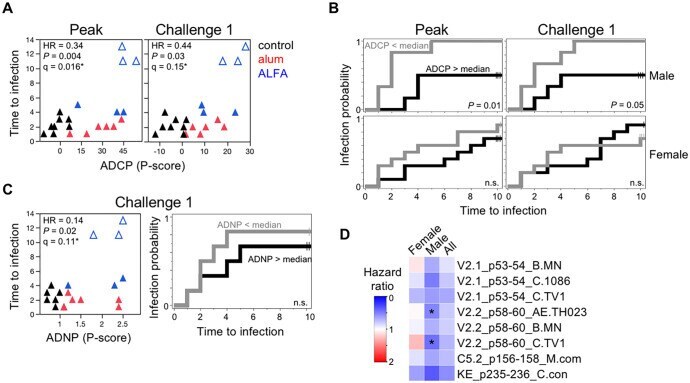
- 10.1371/journal.ppat.1008764.g004 Fig 4 Vaccine-mediated immunologic correlates of protection. (A) ADCP phagocytic (P)-scores are plotted against the challenge number that resulted in SHIV infection for male animals. Peak (month 12.5) and time of challenge (month 15) responses are shown. Unadjusted ( P )- and FDR-adjusted (q)-values reflect significance test of the continuous effects of the responses on the risk of infection (*, q
- Submitted by
- Invitrogen Antibodies (provider)
- Main image

- Experimental details
- 10.1371/journal.ppat.1008764.g005 Fig 5 Env-specific antibody-dependent phagocytic responses elicited by liposomal-MPLA-alum adjuvant in humans. (A) Serum ADCP and ADNP responses in humans vaccinated at weeks 0, 8, 24, and 72 with HIV-1 SF-2 gp120 protein adjuvanted with either alum (red) or liposomal-MPL adsorbed to alum (blue) were assessed at weeks 0 (n = 12 and 13, respectively), 26 (n = 13 each arm), and 112 (n = 11 and 9, respectively). (B) Week 26 ADCP and ADNP responses are plotted against week 26 gp120-specific binding IgG endpoint titers with Spearman's rho and P values indicated. (C) ADCP and (D) ADNP responses are stratified by participant sex to compare responses within each adjuvant study arm (left; M, male; F, female) or between arms within each sex (right). (E) gp120 IIIB Env-specific binding IgG endpoint ELISA titers are shown for all participants combined (left), by sex within each vaccine arm (middle), and by arm within each sex (right). Gray bars depict interquartile range; black lines depict medians; P values reflect Wilcoxon rank-sum test.
