Explore
 Validate
 Learn
Learn Flow cytometry
Antibody data
- Antibody Data
- Antigen structure
- References [4]
- Comments [0]
- Validations
- Flow cytometry [1]
Submit
Validation data
Reference
Comment
Report error
- Product number
- 83-0149-41 - Provider product page

- Provider
- Invitrogen Antibodies
- Product name
- Anti-CD14 Monoclonal Antibody (61D3), eVolve 605, eBioscience™
- Antibody type
- Monoclonal
- Antigen
- Other
- Description
- Notice: The eVolve line of antibody conjugates will be discontinued at the end of February, 2018 and replaced with our improved Invitrogen eBioscience Super Bright conjugated antibodies for flow cytometry. Individual eVovle catalog numbers will become unavailable as inventory depletes. This item has a corresponding Super Bright 600 format available please see Product # 63-0168-41. We will continue to offer a selection of Qdot conjugated antibodies. Description: The eBioCB16 monoclonal antibody recognizes CD16 (Fc gamma RIII), the low-affinity receptor for IgG with an apparent molecular weight of 50-80 kDa. CD16 is represented by two similar genes, CD16A (Fc gamma RIIIA), which exists as a hetero-oligomeric polypeptide-anchored form in macrophages and NK cells and CD16B (Fc gamma RIIIB), which exists as a monomeric GPI-anchored form in neutrophils. Furthermore, there are two known polymorphisms of CD16B, NA-1 and NA-2. Individuals homozygous for NA-2 show a lower phagocytic capacity compared with NA-1. CD16 binds IgG in the form of immune complexes and shows preferential binding of IgG1 and IgG3 isotypes and minimal binding of IgG2 and IgG4. Upon IgG binding, both CD16 isoforms initiate signal transduction cascades that lead to a variety of responses including antibody-dependent cell-mediated cytotoxicity (ADCC), phagocytosis, degranulation, and proliferation. Applications Reported: This eBioCB16 (CB16) antibody has been reported for use in flow cytometric analysis. Applications Tested: This eBioCB16 (CB16) antibody has been pre-titrated and tested by flow cytometric analysis of normal human peripheral blood cells. This can be used at 5 µL per test. A test is defined as the amount of antibody that will stain a cell sample in a final volume of 100 µL. Cell number should be determined empirically but can range from 10^5 to 10^8 cells/test. For best results when using antibodies conjugated to eVolve, we recommend a quick spin of the vial before use and avoid touching the bottom of the vial with the pipet tip. Laser/Filter Recommendation: eVolve 605 can be optimally excited with laser lines from 325-405 nm, and emits near 610 nm. We recommend using the violet (405 nm) laser line for excitation and a 595LP with a 605/40 or 610/20 bandpass filter, or equivalent, for detection. Fixation Recommendation: Antibodies conjugated to eVolve are compatible with eBioscience's fixation and permeabilization buffers. Excitation: 350-590 nm; Emission: 605 nm; Laser: Ultraviolet Laser, Violet Laser Storage and handling: For best results when using antibodies conjugated to eVolve, we recommend a quick spin of the vial before use and avoid touching the bottom of the vial with the pipet tip.
- Reactivity
- Human
- Host
- Mouse
- Isotype
- IgG
- Antibody clone number
- 61D3
- Vial size
- 25 Tests
- Concentration
- 5 µl/Test
- Storage
- 4° C, store in dark, DO NOT FREEZE!
Submitted references Cellular metabolism constrains innate immune responses in early human ontogeny.
AURKA Suppresses Leukemic THP-1 Cell Differentiation through Inhibition of the KDM6B Pathway.
Pulmonary sarcoidosis is associated with high-level inducible co-stimulator (ICOS) expression on lung regulatory T cells--possible implications for the ICOS/ICOS-ligand axis in disease course and resolution.
Noncanonical dendritic cell differentiation and survival driven by a bacteremic pathogen.
Kan B, Michalski C, Fu H, Au HHT, Lee K, Marchant EA, Cheng MF, Anderson-Baucum E, Aharoni-Simon M, Tilley P, Mirmira RG, Ross CJ, Luciani DS, Jan E, Lavoie PM
Nature communications 2018 Nov 16;9(1):4822
Nature communications 2018 Nov 16;9(1):4822
AURKA Suppresses Leukemic THP-1 Cell Differentiation through Inhibition of the KDM6B Pathway.
Park JW, Cho H, Oh H, Kim JY, Seo SB
Molecules and cells 2018 May 31;41(5):444-453
Molecules and cells 2018 May 31;41(5):444-453
Pulmonary sarcoidosis is associated with high-level inducible co-stimulator (ICOS) expression on lung regulatory T cells--possible implications for the ICOS/ICOS-ligand axis in disease course and resolution.
Sakthivel P, Grunewald J, Eklund A, Bruder D, Wahlström J
Clinical and experimental immunology 2016 Feb;183(2):294-306
Clinical and experimental immunology 2016 Feb;183(2):294-306
Noncanonical dendritic cell differentiation and survival driven by a bacteremic pathogen.
Miles B, Scisci E, Carrion J, Sabino GJ, Genco CA, Cutler CW
Journal of leukocyte biology 2013 Aug;94(2):281-9
Journal of leukocyte biology 2013 Aug;94(2):281-9
No comments: Submit comment
Supportive validation
- Submitted by
- Invitrogen Antibodies (provider)
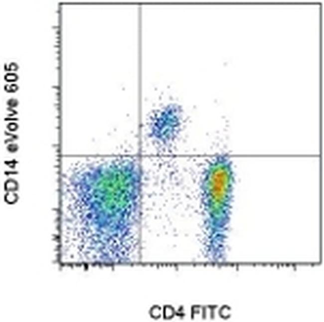
- Main image

- Experimental details
- Staining of normal human peripheral blood cells with Anti-Human CD4 FITC (Product # 11-0049-42) and Anti-Human CD14 eVolve™ 605. Cells in the lymphocyte and monocyte gate were used for analysis.