Explore
 Validate
 Learn
Learn Flow cytometry
Antibody data
- Antibody Data
- Antigen structure
- References [4]
- Comments [0]
- Validations
- Flow cytometry [1]
Submit
Validation data
Reference
Comment
Report error
- Product number
- 83-0149-42 - Provider product page

- Provider
- Invitrogen Antibodies
- Product name
- Anti-CD14 Monoclonal Antibody (61D3), eVolve 605, eBioscience™
- Antibody type
- Monoclonal
- Antigen
- Other
- Description
- Description: The 61D3 monoclonal antibody reacts with human CD14, a 53-55 kDa GPI-linked glycoprotein. CD14 is expressed on monocytes, interfollicular macrophages and some dendritic cells. Complexes of LPS and LBP (LPS-Binding Protein) bind with high affinity to monocytes through the surface CD14. Applications Reported: This 61D3 antibody has been reported for use in flow cytometric analysis. Applications Tested: This 61D3 antibody has been pre-titrated and tested by flow cytometric analysis of normal human peripheral blood cells. This can be used at 5 µL per test. A test is defined as the amount (µg) of antibody that will stain a cell sample in a final volume of 100 µL. Cell number should be determined empirically but can range from 10^5 to 10^8 cells/test. For best results when using antibodies conjugated to eVolve, we recommend a quick spin of the vial before use and avoid touching the bottom of the vial with the pipet tip. Laser/Filter Recommendation: eVolve 605 can be optimally excited with laser lines from 325-405 nm, and emits near 610 nm. We recommend using the violet (405 nm) laser line for excitation and a 595LP with a 605/40 or 610/20 bandpass filter, or equivalent, for detection. Fixation Recommendation: Antibodies conjugated to eVolve are compatible with eBioscience's fixation and permeabilization buffers. Excitation: 350-590 nm; Emission: 605 nm; Laser: Ultraviolet Laser, Violet Laser Storage and handling: For best results when using antibodies conjugated to eVolve, we recommend a quick spin of the vial before use and avoid touching the bottom of the vial with the pipet tip.
- Reactivity
- Human
- Host
- Mouse
- Isotype
- IgG
- Antibody clone number
- 61D3
- Vial size
- 100 Tests
- Concentration
- 5 µl/Test
- Storage
- 4° C, store in dark, DO NOT FREEZE!
Submitted references Cellular metabolism constrains innate immune responses in early human ontogeny.
AURKA Suppresses Leukemic THP-1 Cell Differentiation through Inhibition of the KDM6B Pathway.
Pulmonary sarcoidosis is associated with high-level inducible co-stimulator (ICOS) expression on lung regulatory T cells--possible implications for the ICOS/ICOS-ligand axis in disease course and resolution.
Noncanonical dendritic cell differentiation and survival driven by a bacteremic pathogen.
Kan B, Michalski C, Fu H, Au HHT, Lee K, Marchant EA, Cheng MF, Anderson-Baucum E, Aharoni-Simon M, Tilley P, Mirmira RG, Ross CJ, Luciani DS, Jan E, Lavoie PM
Nature communications 2018 Nov 16;9(1):4822
Nature communications 2018 Nov 16;9(1):4822
AURKA Suppresses Leukemic THP-1 Cell Differentiation through Inhibition of the KDM6B Pathway.
Park JW, Cho H, Oh H, Kim JY, Seo SB
Molecules and cells 2018 May 31;41(5):444-453
Molecules and cells 2018 May 31;41(5):444-453
Pulmonary sarcoidosis is associated with high-level inducible co-stimulator (ICOS) expression on lung regulatory T cells--possible implications for the ICOS/ICOS-ligand axis in disease course and resolution.
Sakthivel P, Grunewald J, Eklund A, Bruder D, Wahlström J
Clinical and experimental immunology 2016 Feb;183(2):294-306
Clinical and experimental immunology 2016 Feb;183(2):294-306
Noncanonical dendritic cell differentiation and survival driven by a bacteremic pathogen.
Miles B, Scisci E, Carrion J, Sabino GJ, Genco CA, Cutler CW
Journal of leukocyte biology 2013 Aug;94(2):281-9
Journal of leukocyte biology 2013 Aug;94(2):281-9
No comments: Submit comment
Supportive validation
- Submitted by
- Invitrogen Antibodies (provider)
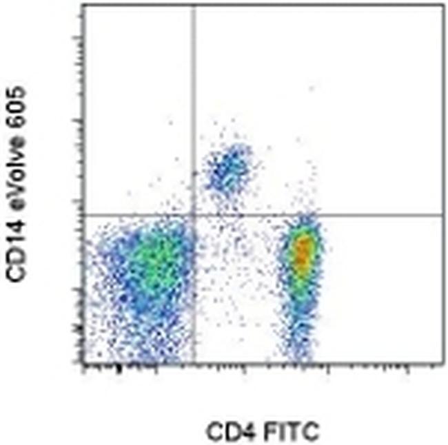
- Main image

- Experimental details
- Staining of normal human peripheral blood cells with Anti-Human CD4 FITC (Product # 11-0049-42) and Anti-Human CD14 eVolve™ 605. Cells in the lymphocyte and monocyte gate were used for analysis.