Explore
 Validate
 Learn
Learn Western blot
 Immunocytochemistry
Antibody data
- Antibody Data
- Antigen structure
- References [14]
- Comments [0]
- Validations
- Immunocytochemistry [1]
- Immunohistochemistry [1]
Submit
Validation data
Reference
Comment
Report error
- Product number
- HPA001887 - Provider product page

- Provider
- Atlas Antibodies
- Proper citation
- Atlas Antibodies Cat#HPA001887, RRID:AB_1078442
- Product name
- Anti-CD14
- Antibody type
- Polyclonal
- Description
- Polyclonal Antibody against Human CD14, Gene description: CD14 molecule, Validated applications: ICC, IHC, WB, Uniprot ID: P08571, Storage: Store at +4°C for short term storage. Long time storage is recommended at -20°C.
- Reactivity
- Human
- Host
- Rabbit
- Conjugate
- Unconjugated
- Isotype
- IgG
- Vial size
- 100 µl
- Concentration
- 0.1 mg/ml
- Storage
- Store at +4°C for short term storage. Long time storage is recommended at -20°C.
- Handling
- The antibody solution should be gently mixed before use.
Submitted references Adipose Tissue Macrophages of the Human Fetus
Spatial mapping reveals granuloma diversity and histopathological superstructure in human tuberculosis
Contribution of Elevated Glucose and Oxidized LDL to Macrophage Inflammation: A Role for PRAS40/Akt-Dependent Shedding of Soluble CD14
A myeloid–stromal niche and gp130 rescue in NOD2-driven Crohn’s disease
RAS mutations drive proliferative chronic myelomonocytic leukemia via a KMT2A-PLK1 axis
Expression of S100A Alarmins in Cord Blood Monocytes Is Highly Associated With Chorioamnionitis and Fetal Inflammation in Preterm Infants.
Identification of novel, immune-mediating extracellular vesicles in human lymphatic effluent draining primary cutaneous melanoma
A High-throughput Bead-based Affinity Assay Enables Analysis of Genital Protein Signatures in Women At Risk of HIV Infection
Levels of human proteins in plasma associated with acute paediatric malaria.
Wingless ligand 5a is a critical regulator of placental growth and survival.
Increased CTLA-4+ T cells and an increased ratio of monocytes with loss of class II (CD14+ HLA-DRlo/neg) found in aggressive pediatric sarcoma patients
LPS and IL‐1 differentially activate mouse and human astrocytes: Role of CD14
High-density lipoprotein endocytosis in endothelial cells
Functional Contribution of Elevated Circulating and Hepatic Non-Classical CD14+CD16+ Monocytes to Inflammation and Human Liver Fibrosis
Radványi Á, Gyurina K, Rácz E, Kovács I, Méhes G, Röszer T
Cells 2024;13(21):1787
Cells 2024;13(21):1787
Spatial mapping reveals granuloma diversity and histopathological superstructure in human tuberculosis
Sawyer A, Patrick E, Edwards J, Wilmott J, Fielder T, Yang Q, Barber D, Ernst J, Britton W, Palendira U, Chen X, Feng C
Journal of Experimental Medicine 2023;220(6)
Journal of Experimental Medicine 2023;220(6)
Contribution of Elevated Glucose and Oxidized LDL to Macrophage Inflammation: A Role for PRAS40/Akt-Dependent Shedding of Soluble CD14
Sanjurjo L, Castelblanco E, Julve J, Villalmanzo N, Téllez É, Ramirez-Morros A, Alonso N, Mauricio D, Sarrias M
Antioxidants 2023;12(5):1083
Antioxidants 2023;12(5):1083
A myeloid–stromal niche and gp130 rescue in NOD2-driven Crohn’s disease
Nayar S, Morrison J, Giri M, Gettler K, Chuang L, Walker L, Ko H, Kenigsberg E, Kugathasan S, Merad M, Chu J, Cho J
Nature 2021;593(7858):275-281
Nature 2021;593(7858):275-281
RAS mutations drive proliferative chronic myelomonocytic leukemia via a KMT2A-PLK1 axis
Carr R, Vorobyev D, Lasho T, Marks D, Tolosa E, Vedder A, Almada L, Yurcheko A, Padioleau I, Alver B, Coltro G, Binder M, Safgren S, Horn I, You X, Solary E, Balasis M, Berger K, Hiebert J, Witzig T, Buradkar A, Graf T, Valent P, Mangaonkar A, Robertson K, Howard M, Kaufmann S, Pin C, Fernandez-Zapico M, Geissler K, Droin N, Padron E, Zhang J, Nikolaev S, Patnaik M
Nature Communications 2021;12(1)
Nature Communications 2021;12(1)
Expression of S100A Alarmins in Cord Blood Monocytes Is Highly Associated With Chorioamnionitis and Fetal Inflammation in Preterm Infants.
Golubinskaya V, Puttonen H, Fyhr IM, Rydbeck H, Hellström A, Jacobsson B, Nilsson H, Mallard C, Sävman K
Frontiers in immunology 2020;11:1194
Frontiers in immunology 2020;11:1194
Identification of novel, immune-mediating extracellular vesicles in human lymphatic effluent draining primary cutaneous melanoma
Maus R, Jakub J, Hieken T, Nevala W, Christensen T, Sutor S, Flotte T, Markovic S
OncoImmunology 2019;8(12):e1667742
OncoImmunology 2019;8(12):e1667742
A High-throughput Bead-based Affinity Assay Enables Analysis of Genital Protein Signatures in Women At Risk of HIV Infection
Månberg A, Bradley F, Qundos U, Guthrie B, Birse K, Noël-Romas L, Lindskog C, Bosire R, Kiarie J, Farquhar C, Burgener A, Nilsson P, Broliden K
Molecular & Cellular Proteomics 2019;18(3):461-476
Molecular & Cellular Proteomics 2019;18(3):461-476
Levels of human proteins in plasma associated with acute paediatric malaria.
Reuterswärd P, Bergström S, Orikiiriza J, Lindquist E, Bergström S, Andersson Svahn H, Ayoglu B, Uhlén M, Wahlgren M, Normark J, Ribacke U, Nilsson P
Malaria journal 2018 Nov 15;17(1):426
Malaria journal 2018 Nov 15;17(1):426
Wingless ligand 5a is a critical regulator of placental growth and survival.
Meinhardt G, Saleh L, Otti GR, Haider S, Velicky P, Fiala C, Pollheimer J, Knöfler M
Scientific reports 2016 Jun 17;6:28127
Scientific reports 2016 Jun 17;6:28127
Increased CTLA-4+ T cells and an increased ratio of monocytes with loss of class II (CD14+ HLA-DRlo/neg) found in aggressive pediatric sarcoma patients
Hingorani P, Maas M, Gustafson M, Dickman P, Adams R, Watanabe M, Eshun F, Williams J, Seidel M, Dietz A
Journal for ImmunoTherapy of Cancer 2015;3(1)
Journal for ImmunoTherapy of Cancer 2015;3(1)
LPS and IL‐1 differentially activate mouse and human astrocytes: Role of CD14
Tarassishin L, Suh H, Lee S
Glia 2014;62(6):999-1013
Glia 2014;62(6):999-1013
High-density lipoprotein endocytosis in endothelial cells
Fruhwürth S, Pavelka M, Bittman R, Kovacs W, Walter K, Röhrl C, Stangl H
World Journal of Biological Chemistry 2013;4(4):131
World Journal of Biological Chemistry 2013;4(4):131
Functional Contribution of Elevated Circulating and Hepatic Non-Classical CD14+CD16+ Monocytes to Inflammation and Human Liver Fibrosis
Bozza P, Zimmermann H, Seidler S, Nattermann J, Gassler N, Hellerbrand C, Zernecke A, Tischendorf J, Luedde T, Weiskirchen R, Trautwein C, Tacke F
PLoS ONE 2010;5(6):e11049
PLoS ONE 2010;5(6):e11049
No comments: Submit comment
Supportive validation
- Submitted by
- Atlas Antibodies (provider)
- Main image

- Experimental details
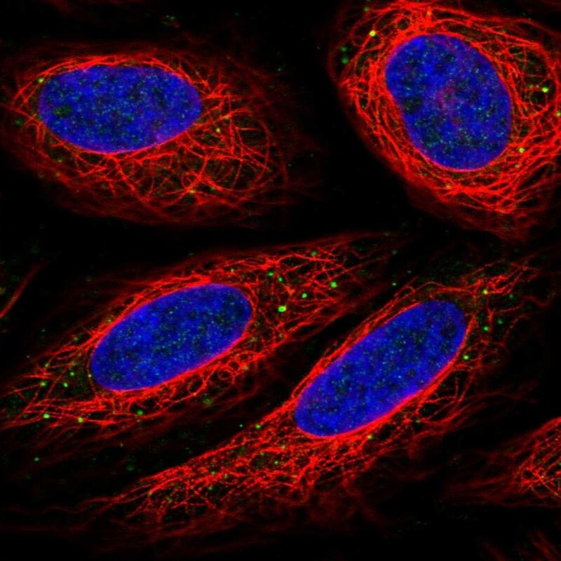
- Immunofluorescent staining of human cell line SiHa shows localization to vesicles.
- Sample type
- Human
Supportive validation
- Submitted by
- Atlas Antibodies (provider)
- Enhanced method
- Orthogonal validation
- Main image

- Experimental details
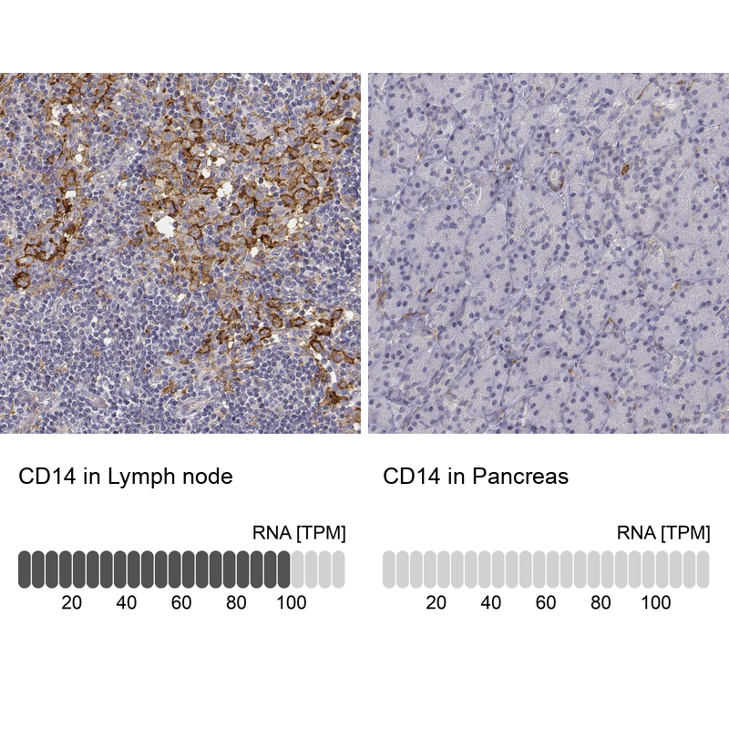
- Immunohistochemistry analysis in human lymph node and pancreas tissues using HPA001887 antibody. Corresponding CD14 RNA-seq data are presented for the same tissues.
- Sample type
- Human
- Protocol
- Protocol