Explore
 Validate
 Learn
Learn Western blot
 Immunohistochemistry
Antibody data
- Antibody Data
- Antigen structure
- References [0]
- Comments [0]
- Validations
- Immunohistochemistry [2]
Submit
Validation data
Reference
Comment
Report error
- Product number
- PA5-22287 - Provider product page

- Provider
- Invitrogen Antibodies
- Product name
- PSMA3 Polyclonal Antibody
- Antibody type
- Polyclonal
- Antigen
- Recombinant full-length protein
- Description
- Recommended positive controls: Molt-4, NIH-3T3, JC, BCL-1. Predicted reactivity: Mouse (94%), Rat (95%), Zebrafish (86%), Xenopus laevis (87%), Chicken (91%), Rhesus Monkey (95%), Bovine (95%). Store product as a concentrated solution. Centrifuge briefly prior to opening the vial.
- Reactivity
- Human, Mouse
- Host
- Rabbit
- Isotype
- IgG
- Vial size
- 100 μL
- Concentration
- 0.36 mg/mL
- Storage
- Store at 4°C short term. For long term storage, store at -20°C, avoiding freeze/thaw cycles.
No comments: Submit comment
Supportive validation
- Submitted by
- Invitrogen Antibodies (provider)
- Main image
- Experimental details
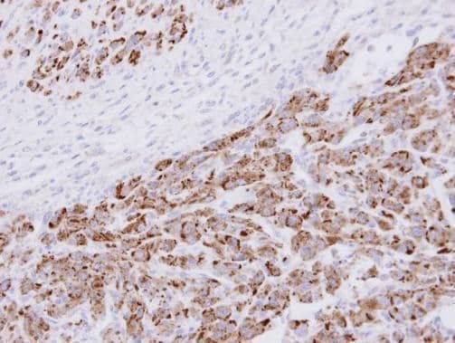
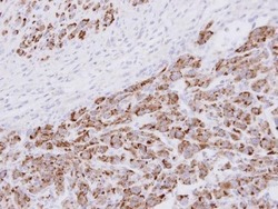
- Immunohistochemistry analysis of PSMA3 was performed on human H1 huES teratoma tissue. To expose target proteins, antigen retrieval was performed using a pressure cooker in 10mM citric acid (pH 6.0). Following antigen retrieval, endogenous peroxidases were blocked with 2.5% hydrogen peroxide. Tissue slide was successively blocked with Avidin D, biotin and protein blocking buffer. Tissues were probed with a PSMA3 polyclonal antibody (Product # PA5-22287) diluted 1:100 overnight at 4°C. After washing tissue slide was incubated with biotinylated anti-rabbit IgG at 37°C for 30 min. After washing tissue slide was incubated at 37°C for 30 min with an HRP kit to conjugate biotinylated anti-rabbit IgG with biotinylated HRP/Avidin. DAB staining for development was performed for 5 min and tissue imaged. Data courtesy of the Antibody Data Exchange Program.

- Submitted by
- Invitrogen Antibodies (provider)
- Main image
- Experimental details
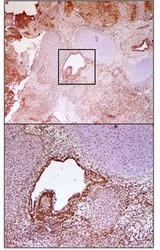
- Immunohistochemical analysis of paraffin-embedded U87 xenograft , using 20S Proteasome alpha3 (Product # PA5-22287) antibody at 1:100 dilution. Antigen Retrieval: EDTA based buffer, pH 8.0, 15 min.