 Explore
 Validate
 Learn
Learn Western blot
Antibody data
- Antibody Data
- Antigen structure
- References [1]
- Comments [0]
- Validations
- Western blot [1]
- Flow cytometry [1]
Submit
Validation data
Reference
Comment
Report error
- Product number
- MAB17161 - Provider product page

- Provider
- Novus Biologicals
- Product name
- Mouse Monoclonal IL-23A/IL-23 P19 Antibody
- Antibody type
- Monoclonal
- Description
- Protein A or G purified from hybridoma culture supernatant. Detects human IL-23 p19 in direct ELISAs and Western blots. In direct ELISAs, approximately 50% cross-reactivity with recombinant human (rh) IL-23 heterodimer is observed, less than 10% cross-reactivity with recombinant mouse (rm) IL-23 heterodimer is observed, and no cross-reactivity with recombinant feline IL-23 p19, recombinant canine (rca) IL-23 p19, and recombinant rat IL-23 p19 is observed.. In Western blots, no cross-reactivity with rcaIL-23 p19 or rmIL-23 p19 is observed.
- Reactivity
- Human
- Host
- Mouse
- Isotype
- IgG
- Vial size
- 100 ug
- Concentration
- LYOPH
- Storage
- Use a manual defrost freezer and avoid repeated freeze-thaw cycles. 12 months from date of receipt, -20 to -70 degreesC as supplied. 1 month, 2 to 8 degreesC under sterile conditions after reconstitution. 6 months, -20 to -70 degreesC under sterile conditions after reconstitution.
Submitted references Induction and activation of human Th17 by targeting antigens to dendritic cells via dectin-1.
Duluc D, Joo H, Ni L, Yin W, Upchurch K, Li D, Xue Y, Klucar P, Zurawski S, Zurawski G, Oh S
Journal of immunology (Baltimore, Md. : 1950) 2014 Jun 15;192(12):5776-88
Journal of immunology (Baltimore, Md. : 1950) 2014 Jun 15;192(12):5776-88
No comments: Submit comment
Supportive validation
- Submitted by
- Novus Biologicals (provider)
- Main image
- Experimental details
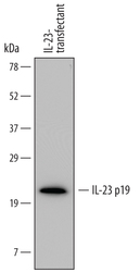
- Detection of Human IL-23 p19 by Western Blot. Western blot shows lysates of CHO Chinese hamster ovary cell line transfected with human IL-23. PVDF membrane was probed with 1 µg/mL of Mouse Anti-Human IL-23 p19 Monoclonal Antibody (Catalog # MAB17161) followed by HRP-conjugated Anti-Mouse IgG Secondary Antibody (Catalog # HAF007). A specific band was detected for IL-23 p19 at approximately 21 kDa (as indicated). This experiment was conducted under reducing conditions and using Immunoblot Buffer Group 1.

Supportive validation
- Submitted by
- Novus Biologicals (provider)
- Main image

- Experimental details
- Detection of IL-23 p19 in Human Blood Monocytes by Flow Cytometry. Human peripheral blood monocytes were treated for 24 hours with 1 μg/mL LPS, then stained with Mouse Anti-Human IL-23 p19 Monoclonal Antibody (Catalog # MAB17161) followed by Allophycocyanin-conjugated Anti-Mouse IgG Secondary Antibody (Catalog # F0101B) and Mouse Anti-Human CD14 PE-conjugated Monoclonal Antibody (Catalog # FAB3832P). Quadrant markers were set based on control antibody staining (Catalog # MAB0041). To facilitate intracellular staining, cells were fixed with paraformaldehyde and permeabilized with saponin.
