Explore
 Validate
 Learn
Learn Western blot
 Immunohistochemistry
Antibody data
- Antibody Data
- Antigen structure
- References [3]
- Comments [0]
- Validations
- Immunohistochemistry [2]
- Other assay [1]
Submit
Validation data
Reference
Comment
Report error
- Product number
- PA5-20239 - Provider product page

- Provider
- Invitrogen Antibodies
- Product name
- IL-23 p19 Polyclonal Antibody
- Antibody type
- Polyclonal
- Antigen
- Synthetic peptide
- Description
- A suggested positive control is mouse pancreas tissue lysate. PA5-20239 can be used with blocking peptide PEP-0358.
- Reactivity
- Human, Mouse
- Host
- Rabbit
- Isotype
- IgG
- Vial size
- 100 μg
- Concentration
- 1 mg/mL
- Storage
- 4°C
Submitted references Primary alterations during the development of hidradenitis suppurativa.
Spontaneous pulmonary hypertension in genetic mouse models of natural killer cell deficiency.
IL23 and TGF-ß diminish macrophage associated metastasis in pancreatic carcinoma.
Dajnoki Z, Somogyi O, Medgyesi B, Jenei A, Szabó L, Gáspár K, Hendrik Z, Gergely P, Imre D, Póliska S, Törőcsik D, Zouboulis CC, Prens EP, Kapitány A, Szegedi A
Journal of the European Academy of Dermatology and Venereology : JEADV 2022 Mar;36(3):462-471
Journal of the European Academy of Dermatology and Venereology : JEADV 2022 Mar;36(3):462-471
Spontaneous pulmonary hypertension in genetic mouse models of natural killer cell deficiency.
Rätsep MT, Moore SD, Jafri S, Mitchell M, Brady HJM, Mandelboim O, Southwood M, Morrell NW, Colucci F, Ormiston ML
American journal of physiology. Lung cellular and molecular physiology 2018 Dec 1;315(6):L977-L990
American journal of physiology. Lung cellular and molecular physiology 2018 Dec 1;315(6):L977-L990
IL23 and TGF-ß diminish macrophage associated metastasis in pancreatic carcinoma.
Hussain SM, Reed LF, Krasnick BA, Miranda-Carboni G, Fields RC, Bi Y, Elahi A, Ajidahun A, Dickson PV, Deneve JL, Hawkins WG, Shibata D, Glazer ES
Scientific reports 2018 Apr 11;8(1):5808
Scientific reports 2018 Apr 11;8(1):5808
No comments: Submit comment
Supportive validation
- Submitted by
- Invitrogen Antibodies (provider)
- Main image
- Experimental details
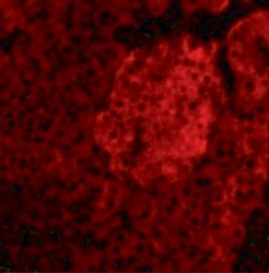
- Immunofluorescent analysis of 4% paraformaldehyde-fixed mouse pancreas tissue labeling IL-23 with IL-23 p19 Polyclonal Antibody (Product # PA5-20239) at 20 µg/mL, followed by goat anti-rabbit IgG secondary antibody at 1:500 dilution (red).

- Submitted by
- Invitrogen Antibodies (provider)
- Main image
- Experimental details
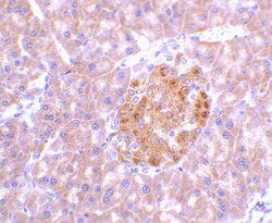
- Immunohistochemical analysis of paraffin-embedded mouse pancreas tissue using IL-23 p19 Polyclonal Antibody (Product # PA5-20239) at 2 µg/mL. Tissue was fixed with formaldehyde and blocked with 0.1 serum for 1 h at RT; antigen retrieval was by heat mediation with a citrate buffer (pH6). Samples were incubated with primary antibody overnight at 4˚ C. A goat anti-rabbit IgG H&L (HRP) at 1/250 was used as secondary. Counter stained with Hematoxylin.

Supportive validation
- Submitted by
- Invitrogen Antibodies (provider)
- Main image
- Experimental details
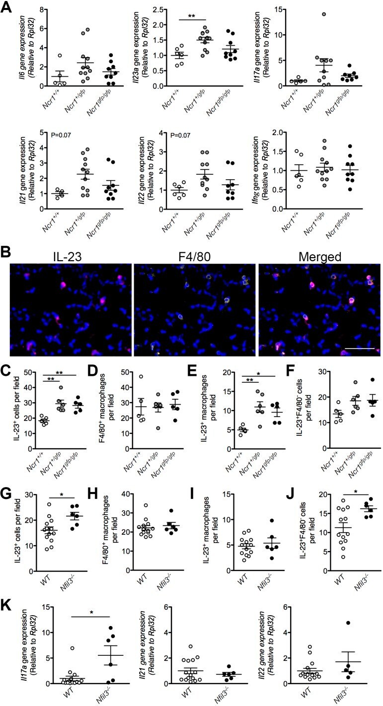
- Fig. 6. Increased IL-23 production and downstream cytokine expression in the Ncr1-Gfp and Nfil3 -/- mouse models of natural killer cell impairment and deficiency. Assessment of inflammatory gene expression in the lungs of Ncr1 +/+ , Ncr1 +/gfp , and Ncr1 gfp/gfp mice ( A ), normalized to the Rpl32 reference gene ( n = 4-11; 1-way ANOVA, Dunnett's posttest vs. Ncr1 +/+ controls). Representative immunofluorescent staining ( B ) of mouse lung tissue for IL-23 (magenta), macrophages (F4/80, yellow), and nuclei (DAPI, blue). Scale bar = 50 mum. Quantification of IL-23 + cells ( C and G ), F4/80 + macrophages ( D and H ), IL-23 + macrophages ( E and I ), and IL-23 + F4/80 - cells ( F and J ) in the lungs of both Ncr1-Gfp mice ( C - F ) ( n = 5-6; 1-way ANOVA, Dunnett's posttest vs. Ncr1 +/+ controls) and Nfil3 -/- mice ( G - J ) with pulmonary hypertension or WT littermate controls ( n = 6-13; Student's t -test). Expression of Th17-associated cytokines ( K ) in the lungs of Nfil3 -/- mice with pulmonary hypertension and WT littermate controls ( n = 5-13; Student's t -test). * P < 0.05 and ** P < 0.01. Means +- SE.