 Explore
 Validate
 Learn
Learn13-8500
antibody from Invitrogen Antibodies
Targeting: JUP
CTNNG, DP3, DPIII, PDGB, PKGB
 Western blot
 Immunocytochemistry
 Immunoprecipitation Immunohistochemistry Flow cytometry Other assay
Antibody data
- Antibody Data
- Antigen structure
- References [26]
- Comments [0]
- Validations
- Immunocytochemistry [2]
- Flow cytometry [1]
- Other assay [17]
Submit
Validation data
Reference
Comment
Report error
- Product number
- 13-8500 - Provider product page

- Provider
- Invitrogen Antibodies
- Product name
- gamma Catenin Monoclonal Antibody (PG-11E4)
- Antibody type
- Monoclonal
- Antigen
- Other
- Reactivity
- Human, Mouse
- Host
- Mouse
- Isotype
- IgG
- Antibody clone number
- PG-11E4
- Vial size
- 100 μg
- Concentration
- 0.5 mg/mL
- Storage
- -20°C
Submitted references Premature Termination Codon in 5' Region of Desmoplakin and Plakoglobin Genes May Escape Nonsense-Mediated Decay through the Reinitiation of Translation.
The fine-tuning of endoplasmic reticulum stress response and autophagy activation during trophoblast syncytialization.
Mechanical loading of desmosomes depends on the magnitude and orientation of external stress.
SuperSILAC Quantitative Proteome Profiling of Murine Middle Ear Epithelial Cell Remodeling with NTHi.
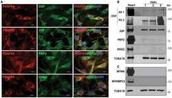
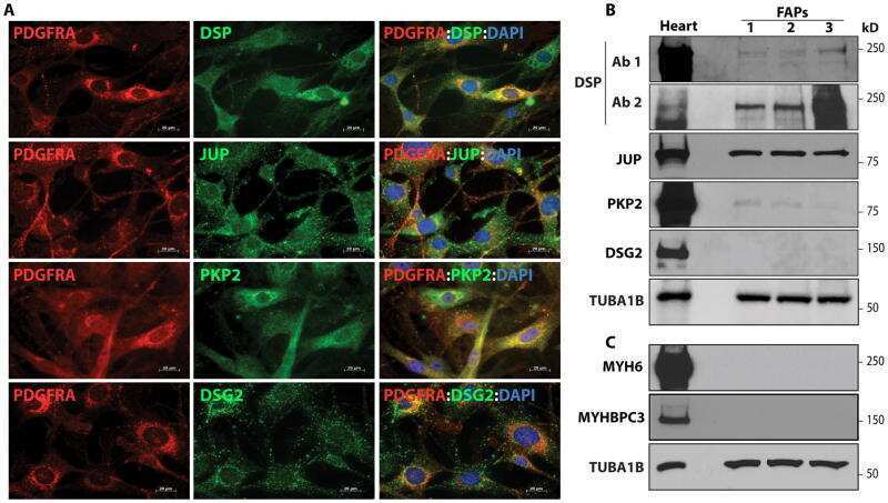
Cardiac Fibro-Adipocyte Progenitors Express Desmosome Proteins and Preferentially Differentiate to Adipocytes Upon Deletion of the Desmoplakin Gene.
Glucocorticoids and histone deacetylase inhibitors cooperate to block the invasiveness of basal-like breast cancer cells through novel mechanisms.
Assessment of desmosomal components (desmoglein 1-3, plakoglobin) in cardia mucosa in relation to gastroesophageal reflux disease and Helicobacter pylori infection.
EpCAM contributes to formation of functional tight junction in the intestinal epithelium by recruiting claudin proteins.
Functional effects of the TMEM43 Ser358Leu mutation in the pathogenesis of arrhythmogenic right ventricular cardiomyopathy.
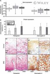
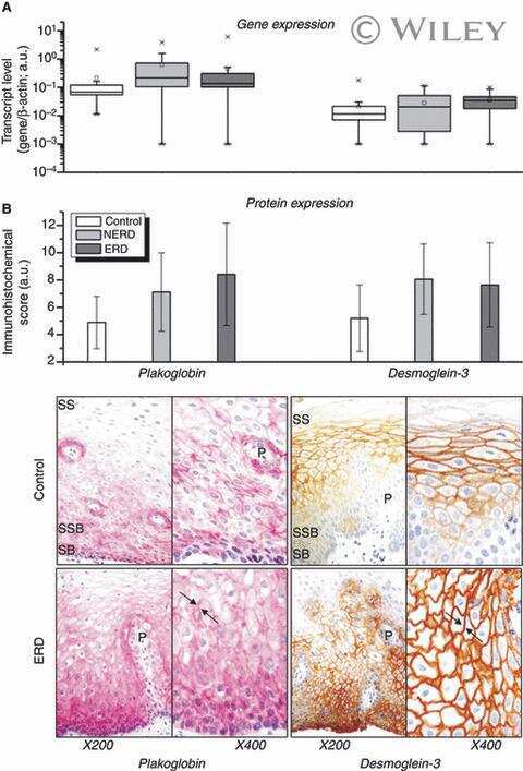
Gastro-oesophageal reflux disease is associated with up-regulation of desmosomal components in oesophageal mucosa.
SOX4 interacts with plakoglobin in a Wnt3a-dependent manner in prostate cancer cells.
Nuclear plakoglobin is essential for differentiation of cardiac progenitor cells to adipocytes in arrhythmogenic right ventricular cardiomyopathy.
Genetic fate mapping identifies second heart field progenitor cells as a source of adipocytes in arrhythmogenic right ventricular cardiomyopathy.
Long-term, multilineage hematopoiesis occurs in the combined absence of beta-catenin and gamma-catenin.
Characterization of a novel type of adherens junction in meningiomas and the derived cell line HBL-52.
Rab4A GTPase catenin interactions are involved in cell junction dynamics in the testis.
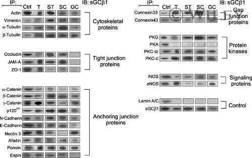
Adjudin-mediated junction restructuring in the seminiferous epithelium leads to displacement of soluble guanylate cyclase from adherens junctions.
The duality of beta-catenin function: a requirement in lens morphogenesis and signaling suppression of lens fate in periocular ectoderm.
Modulating the strength of cadherin adhesion: evidence for a novel adhesion complex.
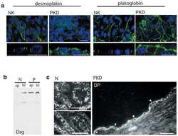
Mispolarization of desmosomal proteins and altered intercellular adhesion in autosomal dominant polycystic kidney disease.
Spink5-deficient mice mimic Netherton syndrome through degradation of desmoglein 1 by epidermal protease hyperactivity.
Identification of a role for beta-catenin in the establishment of a bipolar mitotic spindle.
C-terminal-binding protein corepresses epithelial and proapoptotic gene expression programs.
Hantavirus infection induces the expression of RANTES and IP-10 without causing increased permeability in human lung microvascular endothelial cells.
A comparative evaluation of beta-catenin and plakoglobin signaling activity.
Tumor-derived mutated E-cadherin influences beta-catenin localization and increases susceptibility to actin cytoskeletal changes induced by pervanadate.
Vallverdú-Prats M, Brugada R, Alcalde M
International journal of molecular sciences 2022 Jan 7;23(2)
International journal of molecular sciences 2022 Jan 7;23(2)
The fine-tuning of endoplasmic reticulum stress response and autophagy activation during trophoblast syncytialization.
Bastida-Ruiz D, Yart L, Wuillemin C, Ribaux P, Morris N, Epiney M, Martinez de Tejada B, Cohen M
Cell death & disease 2019 Sep 9;10(9):651
Cell death & disease 2019 Sep 9;10(9):651
Mechanical loading of desmosomes depends on the magnitude and orientation of external stress.
Price AJ, Cost AL, Ungewiß H, Waschke J, Dunn AR, Grashoff C
Nature communications 2018 Dec 11;9(1):5284
Nature communications 2018 Dec 11;9(1):5284
SuperSILAC Quantitative Proteome Profiling of Murine Middle Ear Epithelial Cell Remodeling with NTHi.
Val S, Burgett K, Brown KJ, Preciado D
PloS one 2016;11(2):e0148612
PloS one 2016;11(2):e0148612
Cardiac Fibro-Adipocyte Progenitors Express Desmosome Proteins and Preferentially Differentiate to Adipocytes Upon Deletion of the Desmoplakin Gene.
Lombardi R, Chen SN, Ruggiero A, Gurha P, Czernuszewicz GZ, Willerson JT, Marian AJ
Circulation research 2016 Jun 24;119(1):41-54
Circulation research 2016 Jun 24;119(1):41-54
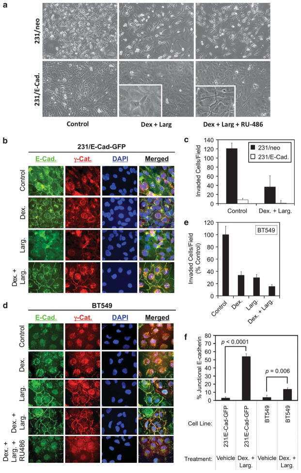
Glucocorticoids and histone deacetylase inhibitors cooperate to block the invasiveness of basal-like breast cancer cells through novel mechanisms.
Law ME, Corsino PE, Jahn SC, Davis BJ, Chen S, Patel B, Pham K, Lu J, Sheppard B, Nørgaard P, Hong J, Higgins P, Kim JS, Luesch H, Law BK
Oncogene 2013 Mar 7;32(10):1316-29
Oncogene 2013 Mar 7;32(10):1316-29
Assessment of desmosomal components (desmoglein 1-3, plakoglobin) in cardia mucosa in relation to gastroesophageal reflux disease and Helicobacter pylori infection.
Wex T, Kuester D, Mönkemüller K, Stahr A, Fry LC, Kandulski A, Kropf S, Roessner A, Malfertheiner P
Human pathology 2012 Oct;43(10):1745-54
Human pathology 2012 Oct;43(10):1745-54
EpCAM contributes to formation of functional tight junction in the intestinal epithelium by recruiting claudin proteins.
Lei Z, Maeda T, Tamura A, Nakamura T, Yamazaki Y, Shiratori H, Yashiro K, Tsukita S, Hamada H
Developmental biology 2012 Nov 15;371(2):136-45
Developmental biology 2012 Nov 15;371(2):136-45
Functional effects of the TMEM43 Ser358Leu mutation in the pathogenesis of arrhythmogenic right ventricular cardiomyopathy.
Rajkumar R, Sembrat JC, McDonough B, Seidman CE, Ahmad F
BMC medical genetics 2012 Mar 29;13:21
BMC medical genetics 2012 Mar 29;13:21
Gastro-oesophageal reflux disease is associated with up-regulation of desmosomal components in oesophageal mucosa.
Wex T, Mönkemüller K, Stahr A, Kuester D, Fry LC, Völkel S, Kandulski A, Roessner A, Malfertheiner P
Histopathology 2012 Feb;60(3):405-15
Histopathology 2012 Feb;60(3):405-15
SOX4 interacts with plakoglobin in a Wnt3a-dependent manner in prostate cancer cells.
Lai YH, Cheng J, Cheng D, Feasel ME, Beste KD, Peng J, Nusrat A, Moreno CS
BMC cell biology 2011 Nov 19;12:50
BMC cell biology 2011 Nov 19;12:50
Nuclear plakoglobin is essential for differentiation of cardiac progenitor cells to adipocytes in arrhythmogenic right ventricular cardiomyopathy.
Lombardi R, da Graca Cabreira-Hansen M, Bell A, Fromm RR, Willerson JT, Marian AJ
Circulation research 2011 Dec 9;109(12):1342-53
Circulation research 2011 Dec 9;109(12):1342-53
Genetic fate mapping identifies second heart field progenitor cells as a source of adipocytes in arrhythmogenic right ventricular cardiomyopathy.
Lombardi R, Dong J, Rodriguez G, Bell A, Leung TK, Schwartz RJ, Willerson JT, Brugada R, Marian AJ
Circulation research 2009 May 8;104(9):1076-84
Circulation research 2009 May 8;104(9):1076-84
Long-term, multilineage hematopoiesis occurs in the combined absence of beta-catenin and gamma-catenin.
Jeannet G, Scheller M, Scarpellino L, Duboux S, Gardiol N, Back J, Kuttler F, Malanchi I, Birchmeier W, Leutz A, Huelsken J, Held W
Blood 2008 Jan 1;111(1):142-9
Blood 2008 Jan 1;111(1):142-9
Characterization of a novel type of adherens junction in meningiomas and the derived cell line HBL-52.
Akat K, Bleck CK, Lee YM, Haselmann-Weiss U, Kartenbeck J
Cell and tissue research 2008 Feb;331(2):401-12
Cell and tissue research 2008 Feb;331(2):401-12
Rab4A GTPase catenin interactions are involved in cell junction dynamics in the testis.
Mruk DD, Lau AS, Sarkar O, Xia W
Journal of andrology 2007 Sep-Oct;28(5):742-54
Journal of andrology 2007 Sep-Oct;28(5):742-54
Adjudin-mediated junction restructuring in the seminiferous epithelium leads to displacement of soluble guanylate cyclase from adherens junctions.
Sarkar O, Xia W, Mruk DD
Journal of cellular physiology 2006 Jul;208(1):175-87
Journal of cellular physiology 2006 Jul;208(1):175-87
The duality of beta-catenin function: a requirement in lens morphogenesis and signaling suppression of lens fate in periocular ectoderm.
Smith AN, Miller LA, Song N, Taketo MM, Lang RA
Developmental biology 2005 Sep 15;285(2):477-89
Developmental biology 2005 Sep 15;285(2):477-89
Modulating the strength of cadherin adhesion: evidence for a novel adhesion complex.
Kim YJ, Sauer C, Testa K, Wahl JK, Svoboda RA, Johnson KR, Wheelock MJ, Knudsen KA
Journal of cell science 2005 Sep 1;118(Pt 17):3883-94
Journal of cell science 2005 Sep 1;118(Pt 17):3883-94
Mispolarization of desmosomal proteins and altered intercellular adhesion in autosomal dominant polycystic kidney disease.
Silberberg M, Charron AJ, Bacallao R, Wandinger-Ness A
American journal of physiology. Renal physiology 2005 Jun;288(6):F1153-63
American journal of physiology. Renal physiology 2005 Jun;288(6):F1153-63
Spink5-deficient mice mimic Netherton syndrome through degradation of desmoglein 1 by epidermal protease hyperactivity.
Descargues P, Deraison C, Bonnart C, Kreft M, Kishibe M, Ishida-Yamamoto A, Elias P, Barrandon Y, Zambruno G, Sonnenberg A, Hovnanian A
Nature genetics 2005 Jan;37(1):56-65
Nature genetics 2005 Jan;37(1):56-65
Identification of a role for beta-catenin in the establishment of a bipolar mitotic spindle.
Kaplan DD, Meigs TE, Kelly P, Casey PJ
The Journal of biological chemistry 2004 Mar 19;279(12):10829-32
The Journal of biological chemistry 2004 Mar 19;279(12):10829-32
C-terminal-binding protein corepresses epithelial and proapoptotic gene expression programs.
Grooteclaes M, Deveraux Q, Hildebrand J, Zhang Q, Goodman RH, Frisch SM
Proceedings of the National Academy of Sciences of the United States of America 2003 Apr 15;100(8):4568-73
Proceedings of the National Academy of Sciences of the United States of America 2003 Apr 15;100(8):4568-73
Hantavirus infection induces the expression of RANTES and IP-10 without causing increased permeability in human lung microvascular endothelial cells.
Sundstrom JB, McMullan LK, Spiropoulou CF, Hooper WC, Ansari AA, Peters CJ, Rollin PE
Journal of virology 2001 Jul;75(13):6070-85
Journal of virology 2001 Jul;75(13):6070-85
A comparative evaluation of beta-catenin and plakoglobin signaling activity.
Williams BO, Barish GD, Klymkowsky MW, Varmus HE
Oncogene 2000 Nov 23;19(50):5720-8
Oncogene 2000 Nov 23;19(50):5720-8
Tumor-derived mutated E-cadherin influences beta-catenin localization and increases susceptibility to actin cytoskeletal changes induced by pervanadate.
Luber B, Candidus S, Handschuh G, Mentele E, Hutzler P, Feller S, Voss J, Höfler H, Becker KF
Cell adhesion and communication 2000 May;7(5):391-408
Cell adhesion and communication 2000 May;7(5):391-408
No comments: Submit comment
Supportive validation
- Submitted by
- Invitrogen Antibodies (provider)
- Main image
- Experimental details
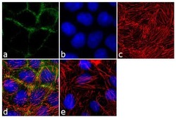
- Immunofluorescence analysis of Gamma-Catenin was performed using 70% confluent log phase CACO2 cells. The cells were fixed with 4% paraformaldehyde for 10 minutes, permeabilized with 0.1% Triton™ X-100 for 10 minutes, and blocked with 1% BSA for 1 hour at room temperature. The cells were labeled with Gamma-Catenin / Plakoglobin (PG-11E4) Mouse Monoclonal Antibody (Product # 13-8500) at 1:250 dilution in0.1% BSA and incubated for 3 hours at room temperature and then labeled with Goat anti-Mouse IgG (H+L) Superclonal™ Secondary Antibody, Alexa Fluor® 488 conjugate (Product # A28175) at a dilution of 1:2000 for 45 minutes at room temperature (Panel a: green). Nuclei (Panel b: blue) were stained with SlowFade® Gold Antifade Mountant with DAPI (Product # S36938). F-actin (Panel c: red) was stained with Rhodamine Phalloidin (Product # R415, 1:300). Panel d represents the merged image showing cytoplasmic localization. Panel e shows the no primary antibody control. The images were captured at 60X magnification.

- Submitted by
- Invitrogen Antibodies (provider)
- Main image

- Experimental details
- Immunofluorescence analysis of Gamma-Catenin was performed using 70% confluent log phase CACO2 cells. The cells were fixed with 4% paraformaldehyde for 10 minutes, permeabilized with 0.1% Triton™ X-100 for 10 minutes, and blocked with 1% BSA for 1 hour at room temperature. The cells were labeled with Gamma-Catenin / Plakoglobin (PG-11E4) Mouse Monoclonal Antibody (Product # 13-8500) at 1:250 dilution in0.1% BSA and incubated for 3 hours at room temperature and then labeled with Goat anti-Mouse IgG (H+L) Superclonal™ Secondary Antibody, Alexa Fluor® 488 conjugate (Product # A28175) at a dilution of 1:2000 for 45 minutes at room temperature (Panel a: green). Nuclei (Panel b: blue) were stained with SlowFade® Gold Antifade Mountant with DAPI (Product # S36938). F-actin (Panel c: red) was stained with Rhodamine Phalloidin (Product # R415, 1:300). Panel d represents the merged image showing cytoplasmic localization. Panel e shows the no primary antibody control. The images were captured at 60X magnification.

Supportive validation
- Submitted by
- Invitrogen Antibodies (provider)
- Main image

- Experimental details
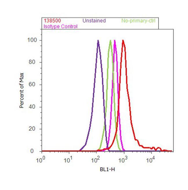
- Flow cytometry analysis of Gamma-Catenin / Plakoglobin was done on Caco-2 cells. Cells were fixed with 70% ethanol for 10 minutes, permeabilized with 0.25% Triton™ X-100 for 20 minutes, and blocked with 5% BSA for 30 minutes at room temperature. Cells were labeled with Gamma-Catenin / Plakoglobin Mouse Monoclonal Antibody (Product # 13-8500, red histogram) or with mouse isotype control (pink histogram) at 3-5 µg/million cells in 2.5% BSA. After incubation at room temperature for 2 hours, the cells were labeled with Alexa Fluor® 488 Rabbit Anti-Mouse Secondary Antibody (Product # A11059) at a dilution of 1:400 for 30 minutes at room temperature. The representative 10,000 cells were acquired and analyzed for each sample using an Attune® Acoustic Focusing Cytometer. The purple histogram represents unstained control cells and the green histogram represents no-primary-antibody control..
Supportive validation
- Submitted by
- Invitrogen Antibodies (provider)
- Main image
- Experimental details
- NULL

- Submitted by
- Invitrogen Antibodies (provider)
- Main image
- Experimental details
- NULL
- Submitted by
- Invitrogen Antibodies (provider)
- Main image
- Experimental details
- NULL
- Submitted by
- Invitrogen Antibodies (provider)
- Main image

- Experimental details
- NULL

- Submitted by
- Invitrogen Antibodies (provider)
- Main image
- Experimental details
- NULL
- Submitted by
- Invitrogen Antibodies (provider)
- Main image
- Experimental details
- NULL

- Submitted by
- Invitrogen Antibodies (provider)
- Main image
- Experimental details
- NULL
- Submitted by
- Invitrogen Antibodies (provider)
- Main image

- Experimental details
- NULL
- Submitted by
- Invitrogen Antibodies (provider)
- Main image
- Experimental details
- NULL
- Submitted by
- Invitrogen Antibodies (provider)
- Main image
- Experimental details
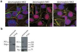
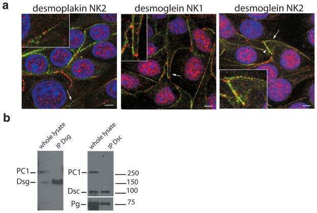
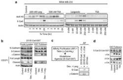
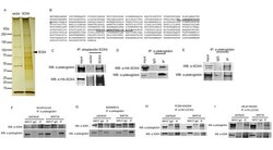
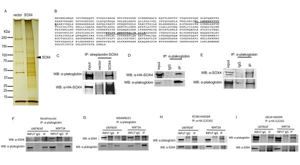
- Figure 1 Plakoglobin binds to SOX4 in LNCaP cells . A . Whole cell lysates prepared from LNCaP cells transfected with pREP4-BLRPwt-HASOX4-IRES-BirA-XL9 or vector only were purified using an streptavidin-magnetic beads, and affinity-purified Vector- and SOX4-complexes were visualized by silver staining. SOX4 protein is indicated by an arrow. The molecular weight markers are as shown on the left. B . Amino acid sequence of plakoglobin with trypsinized peptides from LC-MS/MS analysis indicated in bold and underlined. C , pREP4-BLRPwt-HASOX4-IRES-BirA-XL9 and vector control transfected LNCaP cells. SOX4-associated plakoglobin were analyzed by Western blot with antibodies indicated. Equivalent amounts of the Vector-purified fractions were used to confirm specificity. 5% of transfected LNCaP whole cell lysate was used as Input. D , endogenous plakoglobin IPs were analyzed for transfected SOX4 by Western blot with anti-HA 16B12 mAb. E , endogenous plakoglobin IPs were analyzed for endogenous SOX4 in untransfected LNCaP cells. F, G, endogenous plakoglobin IPs were analyzed for endogenous SOX4 in primary human keratinocytes and breast cancer cell line, MDA-MB-231, respectively. H, I, HASOX4 IPs were analyzed for endogenous plakoglobin in stably-expressed SOX4 PC3M and LNCaP cell lines. F-I, whole cell lysates were harvested and treated with DNase I for 1 hr at room temperature prior to immunoprecipitation.
- Submitted by
- Invitrogen Antibodies (provider)
- Main image
- Experimental details
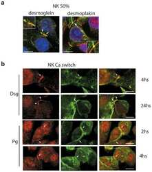
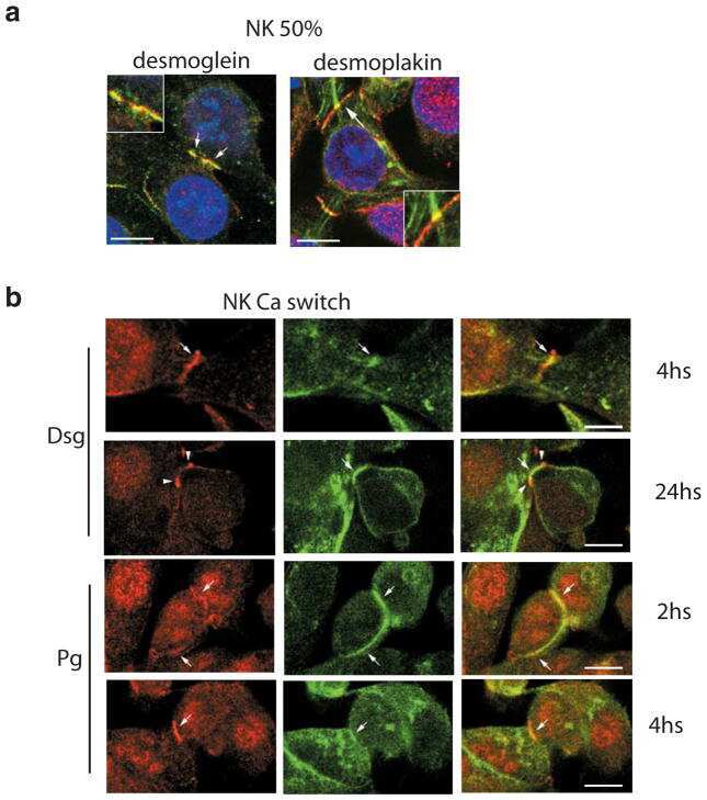
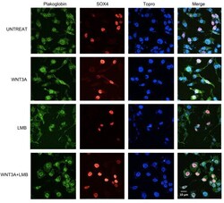
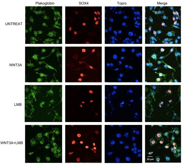
- Figure 2 Wnt signaling induces nuclear colocalization of SOX4 and plakoglobin . Subcellular localization of plakoglobin and SOX4 were examined by confocal microscopy. PC3M HASOX4 stable cell line was treated with 100 ng/ml human recombinant WNT3A or 20 muM leptomycin b (LMB), or both WNT3A+LMB for 24 hr. The fields shown were visualized independently by confocal microscopy at the appropriate wavelength for plakoglobin (488) and SOX4 (543), and Topro (633) respectively, and then the three images were overlaid (Merge). Strong nuclear localization of plakoglobin was observed in the WNT3A+LMB treated cells that expressed HASOX4. Representative fields from these independent repeated experiments are shown. Plakoglobin localizes to the nucleus following WNT3A treatment, and this effect is strongly enhanced by LMB co-treatment, suggesting shuttling of plakoglobin into and out of the nucleus following WNT3A stimulation. Note that plakoglobin nuclear localization is much stronger in cells expressing HASOX4, suggesting SOX4 may facilitate plakoglobin nuclear import.
- Submitted by
- Invitrogen Antibodies (provider)
- Main image
- Experimental details
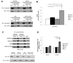
- Figure 3 Interaction between SOX4 and plakoglobin is affected by Wnt3A signaling . A . LNCaP HASOX4 stable cell lines were treated with vehicle, Wnt3A, LMB, or Wnt3A + LMB and anti-HA immunoprecipitations were immunoblotted for the presence of plakoglobin. B , Quantitative analysis of co-IP and Western blots from panel A using the Odyssey (r) Infrared Imaging System (Li-Cor Bioscience). Fold changes were calculated as the ratio of immunoprecipitated plakoglobin to SOX4 relative to the vehicle control panel (untreat). (n = 3, independent biological replicates performed on separate days; error bars represent SEM; *, p < 0.05; **, p < 0.01 for a one-sided paired t-test) C . (Upper) LNCaP HASOX4 stable cell lines were treated with vehicle, WNT3A, LMB, or WNT3A + LMB and anti-HA immunoprecipitations were performed using separate cytosolic and nuclear fractions. (Lower) Anti-AKT and anti-Lamin A antibodies were used as controls to examine markers of cytosolic and nuclear fractions, respectively, to confirm the purity of each fraction. D . Quantitative analysis of co-IP and Western blots from panel C.

- Submitted by
- Invitrogen Antibodies (provider)
- Main image
- Experimental details
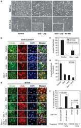
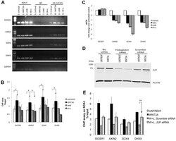
- Figure 4 SOX4 transcriptional activity is modulated by Wnt-induced interaction with plakoglobin . A . ChIP assay of HA-SOX4 bound to the predicted SOX4-binding sites on DICER1 , SOX4 , DHX9 , and AXIN2 promoters. GAPDH is shown as a negative control. B . Quantitative results of ChIP by realtime-PCR. SOX4 promoter occupancy is increased with Wnt treatment, but is strongly reduced by combined WNT3A+LMB treatment. (n = 4; error bars represent SEM; *, p < 0.05 for a one-sided paired t-test) C . SOX4 targets and Wnt downstream genes are inhibited after Wnt induction and LMB treatment. Realtime-PCR expression analysis of SOX4 direct targets and Wnt signaling downstream genes after 24 hrs of WNT3A and LMB treatment. D . siRNA directed against plakoglobin downregulates protein expression in LNCaP HA-SOX4 cell lines. Cells were harvested 48-hr post-transfection and Western blot were probed with anti-plakoglobin and anti-beta-actin antibodies in the presence or absence of WNT3A treatment. E . Quantitative ChIP-qPCR assay of HA-SOX4 following plakoglobin knockdown. HA-SOX4 promoter occupancy is strongly reduced by combined WNT3A and LMB treatment but is partially restored when plakoglobin is knocked down by siRNA treatment. Control scrambled siRNA had no effect (n = 3; error bars represent SEM; *, p < 0.05 for a one-sided paired t-test).

- Submitted by
- Invitrogen Antibodies (provider)
- Main image
- Experimental details
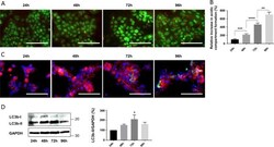
- Fig. 3 Activation of autophagy during syncytialization. a Primary trophoblastic cells were labeled with acridine orange 24, 48, 72 and 96 h after seeding and observed by fluorescence microscopy. Scale bar represents 100 mum. b Acridine orange quantification of the intensity and area of red signal, normalized to the total number of nuclei. n = 4. Data represented as mean +- SEM. ** P

- Submitted by
- Invitrogen Antibodies (provider)
- Main image
- Experimental details
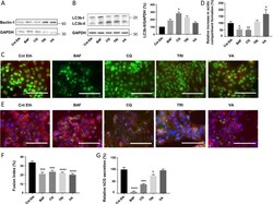
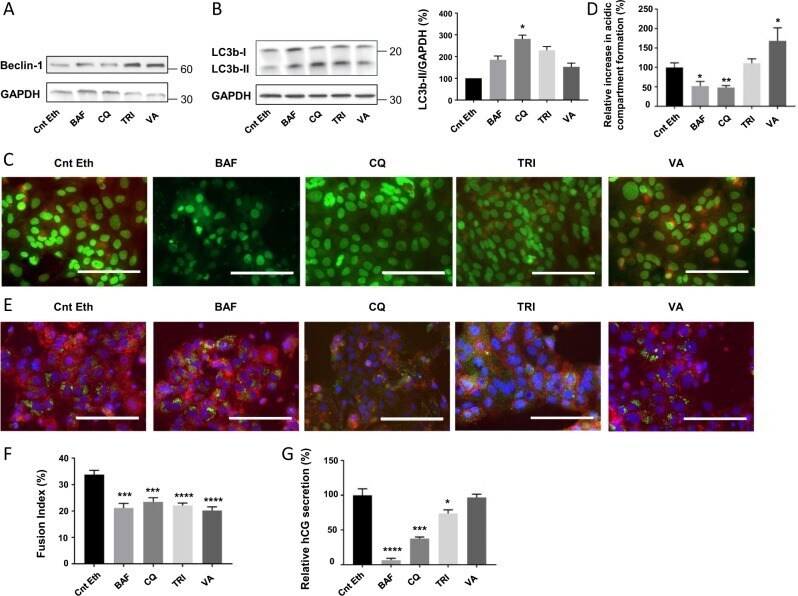
- Fig. 4 Consequences of autophagy modulation with autophagy inhibitors or activators on trophoblastic cell fusion and differentiation. vCTB were purified from human term placenta and seeded for 24 h prior treatment with 10 nM Bafilomycin A1 (BAF), 2 uM Chloroquine (CQ), 10 nM Trichostatin A (TRI), 200 uM Valproic acid (VA), or Ethanol (Control Ethanol, Cnt Eth) for 48 h. a , b Western blotting was performed on the cells. LC3b-II and GAPDH levels were quantified using the ImageJ software, and data are expressed as the fold change relative to the control. n = 4. Data represented as mean +- SEM. * P
- Submitted by
- Invitrogen Antibodies (provider)
- Main image

- Experimental details
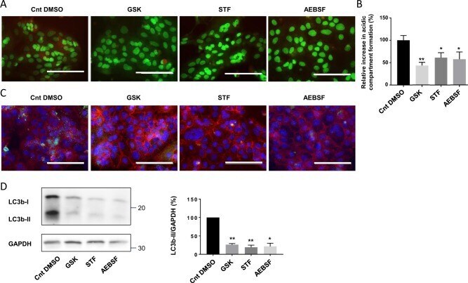
- Fig. 6 Effect of UPR modulation on autophagy activation in trophoblastic cells. vCTB were purified from human term placenta and seeded for 24 h prior treatment with 100 nM GSK2656157 (GSK), 100 uM STF-083010 (STF), 200 uM 4-(2-aminoethyl)benzenesulfonyl fluoride hydrochloride (AEBSF), or DMSO (Control DMSO, Cnt DMSO) for 48 h. a Cells were labeled with acridine orange 48 h post-treatment and observed by fluorescence microscopy. Scale bar represents 100 mum. b Acridine orange quantification of the intensity and area of red signal, normalized to the total number of nuclei. n = 4. Data represented as mean +- SEM. * P
- Submitted by
- Invitrogen Antibodies (provider)
- Main image
- Experimental details
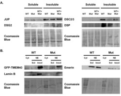
- Figure 2 Solubility assays . Solubility patterns for (A) desmosomal and (B) nuclear envelope proteins were assessed by immunoblots for soluble and insoluble protein fractions extracted from COS-7 cells transfected with wildtype (WT) TMEM43 , mutant (Mut) TMEM43 , or a 1:2 ratio of wildtype to mutant (WT + Mut) TMEM43 . Sol, soluble fraction containing cytoplasmic (Cyt) proteins; Insol, insoluble fraction containing membrane bound proteins. No differences in solubility patterns were found, suggesting that the presence of mutant TMEM43 did not alter the stability of TMEM43 or of other desmosomal or nuclear envelope proteins.
