 Explore
 Validate
 Learn
Learn Western blot
Antibody data
- Antibody Data
- Antigen structure
- References [0]
- Comments [0]
- Validations
- Western blot [5]
- Immunocytochemistry [2]
- Immunohistochemistry [2]
Submit
Validation data
Reference
Comment
Report error
- Product number
- MA5-15905 - Provider product page

- Provider
- Invitrogen Antibodies
- Product name
- gamma Catenin Monoclonal Antibody (4C12)
- Antibody type
- Monoclonal
- Antigen
- Purifed from natural sources
- Description
- MA5-15905 targets JUP in IF, IHC, and WB applications and shows reactivity with Human samples.
- Antibody clone number
- 4C12
- Concentration
- Conc. Not Determined
No comments: Submit comment
Supportive validation
- Submitted by
- Invitrogen Antibodies (provider)
- Main image

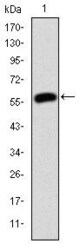
- Experimental details
- Western blot analysis of JUP using a JUP monoclonal antibody (Product # MA5-15905) against a human JUP (AA: 534-740) recombinant protein.

- Submitted by
- Invitrogen Antibodies (provider)
- Main image
- Experimental details
- Western blot analysis of JUP using a JUP monoclonal antibody (Product # MA5-15905) against a human JUP (AA: 534-740) recombinant protein.

- Submitted by
- Invitrogen Antibodies (provider)
- Main image
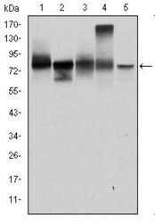
- Experimental details
- Western blot analysis of JUP using JUP monoclonal antibody (Product # MA5-15905) in T47D (1), MCF-7 (2), SKBR-3 (3), A431 (4) and HEK293 (5) cell lysate.
- Submitted by
- Invitrogen Antibodies (provider)
- Main image
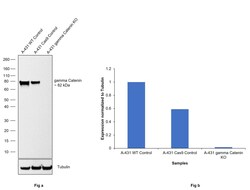
- Experimental details
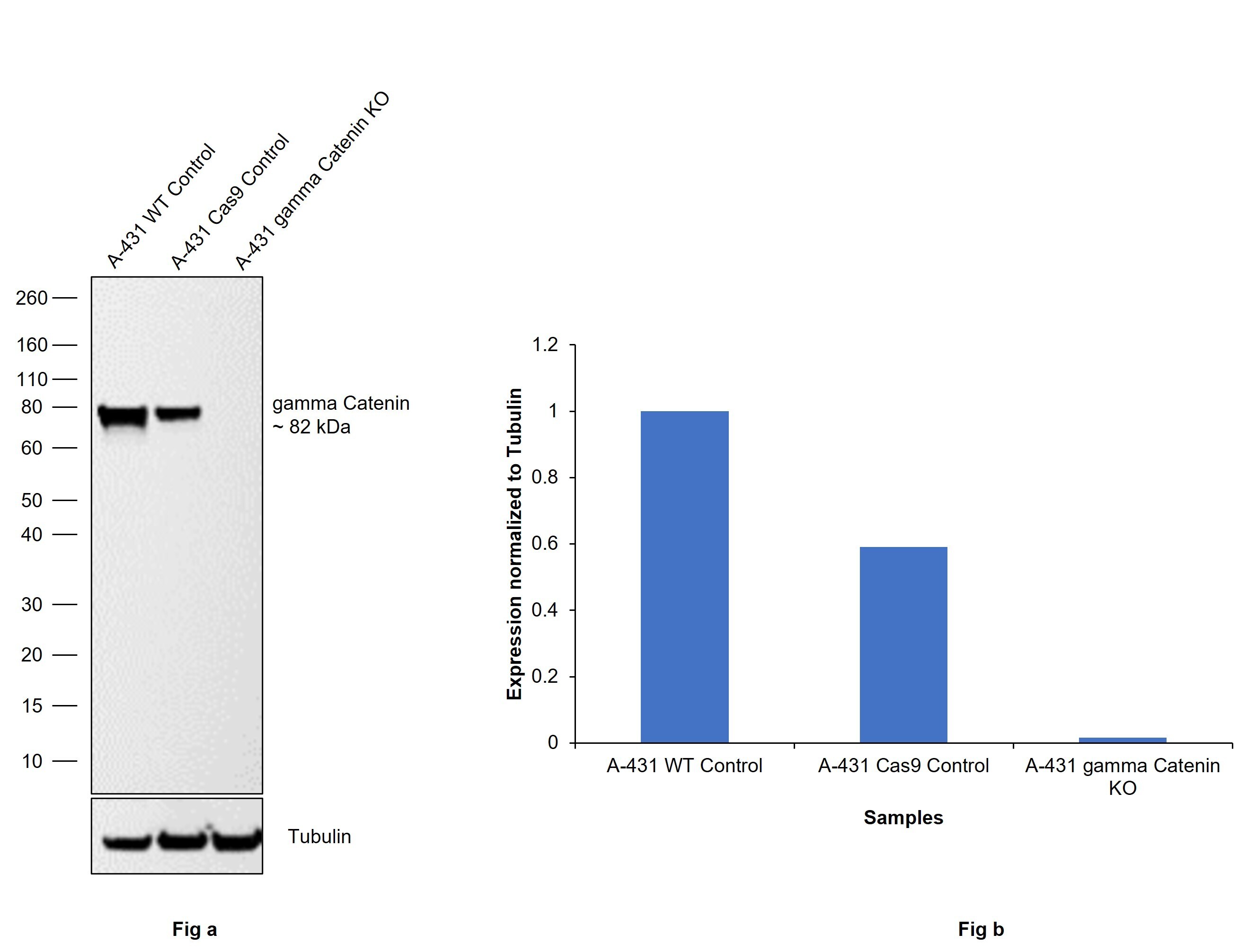
- Knockout of gamma Catenin was achieved by CRISPR-Cas9 genome editing using LentiArray™ Lentiviral sgRNA (Product # A32042, Assay ID CRISPR680765_LV) and LentiArray Cas9 Lentivirus (Product # A32064). Western blot analysis of gamma Catenin was performed by loading 30 µg of A-431 Wild type (Lane 1), A-431 Cas9 (Lane 2) andA-431 gamma Catenin KO (Lane 3) whole cell extracts. The samples were electrophoresed using NuPAGE™ Novex™ 4-12% Bis-Tris Protein Gel (Product # NP0321BOX). Resolved proteins were then transferred onto a nitrocellulose membrane (Product # IB23001) by iBlot® 2 Dry Blotting System (Product # IB21001). The blot was probed with gamma Catenin Monoclonal Antibody (4C12) (Product # MA5-15905, 1:1000 dilution) and Goat anti-Mouse IgG (H+L) Superclonal™ Recombinant Secondary Antibody, HRP (Product # A28177, 1:10,000 dilution) using the iBright™ FL 1500 (Product # A44115). Chemiluminescent detection was performed using SuperSignal™ West Dura Extended Duration Substrate (Product # 34076). Loss of signal upon CRISPR mediated knockout (KO) using the LentiArray™ CRISPR product line confirms that antibody is specific to gamma Catenin.
- Submitted by
- Invitrogen Antibodies (provider)
- Main image
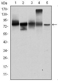
- Experimental details
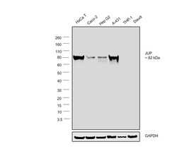
- Western blot was performed using Anti-gamma Catenin Monoclonal Antibody (4C12)(Product # MA5-15905) and a 82kDa band corresponding to gamma Catenin was observed across the cell lines tested except THP-1 and Daudi. Whole cell extracts (30 µg lysate) of HaCaT (Lane 1), Caco-2 (Lane 2), Hep G2 (Lane 3), A-431 (Lane 4), THP-1 (Lane 5) and Daudi (Lane 6) were electrophoresed using NuPAGE™ 4-12% Bis-Tris Protein Gel (Product # NP0321BOX). Resolved proteins were then transferred onto a Nitrocellulose membrane (Product # LC2001) by iBlot® 2 Dry Blotting System (Product # IB21001). The blot was probed with the primary antibody (1:1000 dilution) and detected by chemiluminescence with Goat anti-Mouse IgG (H+L) Superclonal™ Recombinant Secondary Antibody, HRP (Product # A28177,1:4000 dilution) using the iBright FL 1000 (Product # A32752). Chemiluminescent detection was performed using Novex® ECL Chemiluminescent Substrate Reagent Kit (Product # WP20005).

Supportive validation
- Submitted by
- Invitrogen Antibodies (provider)
- Main image

- Experimental details
- Immunofluorescence analysis of U251 cells using JUP monoclonal antibody (Product # MA5-15905) (Green). Blue: DRAQ5 fluorescent DNA dye.

- Submitted by
- Invitrogen Antibodies (provider)
- Main image

- Experimental details
- Immunofluorescence analysis of gamma Catenin was performed using 70% confluent log phase HaCaT cells. The cells were fixed with 4% paraformaldehyde for 15 minutes, permeabilized with 0.1% Triton™ X-100 for 10 minutes, and blocked with 2% BSA for 45 minutes at room temperature. The cells were labeled with gamma Catenin Monoclonal Antibody (4C12) (Product # MA5-15905) at 1:200 dilution in 0.1% BSA, incubated at 4 degree celsius overnight and then labeled with Donkey anti-Mouse IgG (H+L) Highly Cross-Adsorbed Secondary Antibody, Alexa Fluor Plus 488 (Product # A32766), (1:2000 dilution), for 45 minutes at room temperature (Panel a: Green). Nuclei (Panel b:Blue) were stained with SlowFade® Gold Antifade Mountant with DAPI (Product # S36938). F-actin (Panel c: Red) was stained with Rhodamine Phalloidin (Product # R415, 1:300 dilution). Panel d represents the merged image showing cell junction and cytoplasmic localization. Panel e represents control cells with no primary antibody to assess background. The images were captured at 60X magnification.

Supportive validation
- Submitted by
- Invitrogen Antibodies (provider)
- Main image

- Experimental details
- Immunohistochemical analysis of paraffin-embedded rectum tissues using JUP monoclonal antibody (Product # MA5-15905) followed with DAB staining.

- Submitted by
- Invitrogen Antibodies (provider)
- Main image

- Experimental details
- Immunohistochemical analysis of paraffin-embedded stomach cancer tissues using JUP monoclonal antibody (Product # MA5-15905) followed with DAB staining.
