Explore
 Validate
 Learn
Learn Western blot
 Immunocytochemistry
Antibody data
- Antibody Data
- Antigen structure
- References [1]
- Comments [0]
- Validations
- Immunocytochemistry [2]
- Immunoprecipitation [1]
- Immunohistochemistry [2]
- Other assay [1]
Submit
Validation data
Reference
Comment
Report error
- Product number
- PA5-29930 - Provider product page

- Provider
- Invitrogen Antibodies
- Product name
- gamma Catenin Polyclonal Antibody
- Antibody type
- Polyclonal
- Antigen
- Recombinant full-length protein
- Description
- Recommended positive controls: A431. Predicted reactivity: Mouse (99%), Rat (99%), Xenopus laevis (87%), Dog (99%), Pig (98%), Chicken (97%), Rhesus Monkey (100%), Bovine (98%). Store product as a concentrated solution. Centrifuge briefly prior to opening the vial.
- Reactivity
- Human, Mouse
- Host
- Rabbit
- Isotype
- IgG
- Vial size
- 100 μL
- Concentration
- 0.36 mg/mL
- Storage
- Store at 4°C short term. For long term storage, store at -20°C, avoiding freeze/thaw cycles.
Submitted references Central role for GSK3β in the pathogenesis of arrhythmogenic cardiomyopathy.
Chelko SP, Asimaki A, Andersen P, Bedja D, Amat-Alarcon N, DeMazumder D, Jasti R, MacRae CA, Leber R, Kleber AG, Saffitz JE, Judge DP
JCI insight 2016 Apr 21;1(5)
JCI insight 2016 Apr 21;1(5)
No comments: Submit comment
Supportive validation
- Submitted by
- Invitrogen Antibodies (provider)
- Main image
- Experimental details
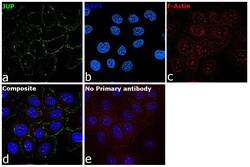
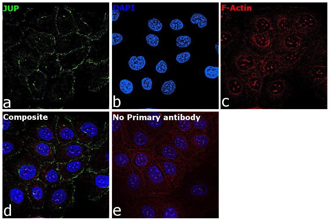
- Immunofluorescence analysis of gamma Catenin was performed using 70% confluent log phase HaCaT cells. The cells were fixed with 4% paraformaldehyde for 10 minutes, permeabilized with 0.1% Triton™ X-100 for 10 minutes, and blocked with 2% BSA for 45 minutes at room temperature. The cells were labeled with gamma Catenin Polyclonal Antibody (Product # PA5-29930) at 1:200 dilution in 0.1% BSA, incubated at 4 degree celsius overnight and then labeled with Donkey anti-Rabbit IgG (H+L) Highly Cross-Adsorbed Secondary Antibody, Alexa Fluor Plus 488 (Product # A32790), (1:2000 dilution), for 45 minutes at room temperature (Panel a: Green). Nuclei (Panel b:Blue) were stained with SlowFade® Gold Antifade Mountant with DAPI (Product # S36938). F-actin (Panel c: Red) was stained with Rhodamine Phalloidin (Product # R415, 1:300 dilution). Panel d represents the merged image showing membrane and cytoplasm localization. Panel e represents control cells with no primary antibody to assess background. The images were captured at 60X magnification.

- Submitted by
- Invitrogen Antibodies (provider)
- Main image

- Experimental details
- Immunofluorescence analysis of gamma Catenin was performed using 70% confluent log phase HaCaT cells. The cells were fixed with 4% paraformaldehyde for 10 minutes, permeabilized with 0.1% Triton™ X-100 for 10 minutes, and blocked with 2% BSA for 45 minutes at room temperature. The cells were labeled with gamma Catenin Polyclonal Antibody (Product # PA5-29930) at 1:200 dilution in 0.1% BSA, incubated at 4 degree celsius overnight and then labeled with Donkey anti-Rabbit IgG (H+L) Highly Cross-Adsorbed Secondary Antibody, Alexa Fluor Plus 488 (Product # A32790), (1:2000 dilution), for 45 minutes at room temperature (Panel a: Green). Nuclei (Panel b:Blue) were stained with SlowFade® Gold Antifade Mountant with DAPI (Product # S36938). F-actin (Panel c: Red) was stained with Rhodamine Phalloidin (Product # R415, 1:300 dilution). Panel d represents the merged image showing membrane and cytoplasm localization. Panel e represents control cells with no primary antibody to assess background. The images were captured at 60X magnification.
Supportive validation
- Submitted by
- Invitrogen Antibodies (provider)
- Main image

- Experimental details
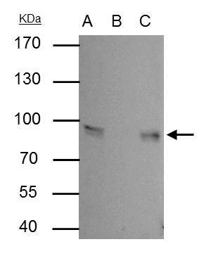
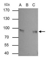
- Gamma Catenin antibody immunoprecipitates Gamma Catenin protein in IP experiments. IP Sample: A431 whole cell lysate/extract A : 30 µg whole cell lysate/extract of Gamma Catenin protein expressing A431 cells B : Control with 2.5 µg of pre-immune rabbit IgG C : Immunoprecipitation of Gamma Catenin by 2.5 µg of Gamma Catenin antibody (Product # PA5-29930) 7.5% SDS-PAGE The immunoprecipitated Gamma Catenin protein was detected by Gamma Catenin antibody (Product # PA5-29930) diluted at 1:1,000. Anti-rabbit IgG (HRP) was used as a secondary reagent.

Supportive validation
- Submitted by
- Invitrogen Antibodies (provider)
- Main image
- Experimental details
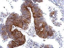
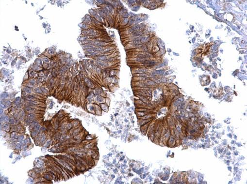
- gamma Catenin Polyclonal Antibody detects gamma Catenin protein at cytosol and membrane on human colon carcinoma by immunohistochemical analysis. Sample: Paraffin-embedded human colon carcinoma. Gamma Catenin Polyclonal Antibody (Product # PA5-29930) dilution: 1:500. Antigen Retrieval: EDTA based buffer, pH 8.0, 15 min.
- Submitted by
- Invitrogen Antibodies (provider)
- Main image
- Experimental details
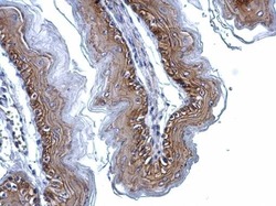
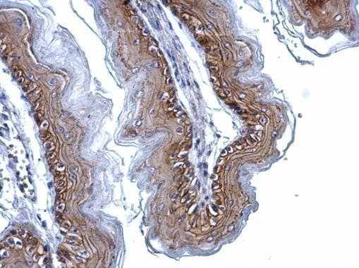
- gamma Catenin Polyclonal Antibody detects gamma Catenin protein at cytosol and membrane on mouse esophagus by immunohistochemical analysis. Sample: Paraffin-embedded mouse esophagus. Gamma Catenin Polyclonal Antibody (Product # PA5-29930) dilution: 1:500. Antigen Retrieval: EDTA based buffer, pH 8.0, 15 min.
Supportive validation
- Submitted by
- Invitrogen Antibodies (provider)
- Main image
- Experimental details
- Gamma Catenin antibody immunoprecipitates Gamma Catenin protein in IP experiments. IP Sample: A431 whole cell lysate/extract A : 30 µg whole cell lysate/extract of Gamma Catenin protein expressing A431 cells B : Control with 2.5 µg of pre-immune rabbit IgG C : Immunoprecipitation of Gamma Catenin by 2.5 µg of Gamma Catenin antibody (Product # PA5-29930) 7.5% SDS-PAGE The immunoprecipitated Gamma Catenin protein was detected by Gamma Catenin antibody (Product # PA5-29930) diluted at 1:1,000. Anti-rabbit IgG (HRP) was used as a secondary reagent.