 Explore
 Validate
 Learn
LearnNB600-1317
antibody from Novus Biologicals
Targeting: CD44
CD44R, CSPG8, HCELL, IN, MC56, MDU2, MDU3, MIC4, Pgp1
 Western blot
 Immunohistochemistry
Antibody data
- Antibody Data
- Antigen structure
- References [15]
- Comments [0]
- Validations
- Immunohistochemistry [1]
- Flow cytometry [2]
Submit
Validation data
Reference
Comment
Report error
- Product number
- NB600-1317 - Provider product page

- Provider
- Novus Biologicals
- Proper citation
- Novus Cat#NB600-1317, RRID:AB_10002839
- Product name
- Mouse Monoclonal CD44 Antibody
- Antibody type
- Monoclonal
- Description
- Protein G purified. This recognizes the rat CD44 cell surface antigen, expressed by T cells, B cells, macrophages and thymocytes.
- Reactivity
- Rat
- Host
- Mouse
- Isotype
- IgG
- Vial size
- 0.1 ml
- Concentration
- 1.0 mg/ml
- Storage
- Store at 4C short term. Aliquot and store at -20C long term. Avoid freeze-thaw cycles.
Submitted references TDP-43 knockdown causes innate immune activation via protein kinase R in astrocytes.
Focal and segmental glomerulosclerosis in murine models: a histological and ultrastructural characterization with immunohistochemistry correlation of glomerular CD44 and WT1 expression.
Pressure-Induced Alterations in PEDF and PEDF-R Expression: Implications for Neuroprotective Signaling in Glaucoma.
Systemic induction of the angiogenesis switch by the tetraspanin D6.1A/CO-029.
Distribution patterns of the membrane glycoprotein CD44 during the foreign-body reaction to a degradable biomaterial in rats and mice.
Functional activity of CD44 isoforms in haemopoiesis of the rat.
Functional activity of CD44 isoforms in haemopoiesis of the rat.
Enrichment of early fetal-liver hemopoietic stem cells of the rat using monoclonal antibodies against the transferrin receptor, Thy-1, and MRC-OX82.
Enrichment of early fetal-liver hemopoietic stem cells of the rat using monoclonal antibodies against the transferrin receptor, Thy-1, and MRC-OX82.
CD44 exhibits a cell type dependent interaction with triton X-100 insoluble, lipid rich, plasma membrane domains.
Monoclonal antibodies to CD44 and their influence on hyaluronan recognition.
Monoclonal antibodies to CD44 and their influence on hyaluronan recognition.
Functional and phenotypical analysis of subsets of rat CD4+ T cells.
Functional and phenotypical analysis of subsets of rat CD4+ T cells.
Prevention of tumor metastasis formation by anti-variant CD44.
LaRocca TJ, Mariani A, Watkins LR, Link CD
Neurobiology of disease 2019 Dec;132:104514
Neurobiology of disease 2019 Dec;132:104514
Focal and segmental glomerulosclerosis in murine models: a histological and ultrastructural characterization with immunohistochemistry correlation of glomerular CD44 and WT1 expression.
Husain S, Ginawi I, Bashir AI, Kfoury H, Al Johani TE, Hagar H, Raddaoui L, Al Ghonaim M, Alsuwaida A
Ultrastructural pathology 2018 Sep-Oct;42(5):430-439
Ultrastructural pathology 2018 Sep-Oct;42(5):430-439
Pressure-Induced Alterations in PEDF and PEDF-R Expression: Implications for Neuroprotective Signaling in Glaucoma.
Lee SJ, Duncan DS, Echevarria FD, McLaughlin WM, Hatcher JB, Sappington RM
Journal of clinical & experimental ophthalmology 2015 Oct;6(5)
Journal of clinical & experimental ophthalmology 2015 Oct;6(5)
Systemic induction of the angiogenesis switch by the tetraspanin D6.1A/CO-029.
Gesierich S, Berezovskiy I, Ryschich E, Zöller M
Cancer research 2006 Jul 15;66(14):7083-94
Cancer research 2006 Jul 15;66(14):7083-94
Distribution patterns of the membrane glycoprotein CD44 during the foreign-body reaction to a degradable biomaterial in rats and mice.
Bonnema H, Popa ER, van Timmeren MM, van Wachem PB, de Leij LF, van Luyn MJ
Journal of biomedical materials research. Part A 2003 Mar 1;64(3):502-8
Journal of biomedical materials research. Part A 2003 Mar 1;64(3):502-8
Functional activity of CD44 isoforms in haemopoiesis of the rat.
Khaldoyanidi S, Schnabel D, Föhr N, Zöller M
British journal of haematology 1997 Jan;96(1):31-45
British journal of haematology 1997 Jan;96(1):31-45
Functional activity of CD44 isoforms in haemopoiesis of the rat.
Khaldoyanidi S, Schnabel D, Föhr N, Zöller M
British journal of haematology 1997 Jan;96(1):31-45
British journal of haematology 1997 Jan;96(1):31-45
Enrichment of early fetal-liver hemopoietic stem cells of the rat using monoclonal antibodies against the transferrin receptor, Thy-1, and MRC-OX82.
Crook K, Hunt SV
Developmental immunology 1996;4(4):235-46
Developmental immunology 1996;4(4):235-46
Enrichment of early fetal-liver hemopoietic stem cells of the rat using monoclonal antibodies against the transferrin receptor, Thy-1, and MRC-OX82.
Crook K, Hunt SV
Developmental immunology 1996;4(4):235-46
Developmental immunology 1996;4(4):235-46
CD44 exhibits a cell type dependent interaction with triton X-100 insoluble, lipid rich, plasma membrane domains.
Neame SJ, Uff CR, Sheikh H, Wheatley SC, Isacke CM
Journal of cell science 1995 Sep;108 ( Pt 9):3127-35
Journal of cell science 1995 Sep;108 ( Pt 9):3127-35
Monoclonal antibodies to CD44 and their influence on hyaluronan recognition.
Zheng Z, Katoh S, He Q, Oritani K, Miyake K, Lesley J, Hyman R, Hamik A, Parkhouse RM, Farr AG, Kincade PW
The Journal of cell biology 1995 Jul;130(2):485-95
The Journal of cell biology 1995 Jul;130(2):485-95
Monoclonal antibodies to CD44 and their influence on hyaluronan recognition.
Zheng Z, Katoh S, He Q, Oritani K, Miyake K, Lesley J, Hyman R, Hamik A, Parkhouse RM, Farr AG, Kincade PW
The Journal of cell biology 1995 Jul;130(2):485-95
The Journal of cell biology 1995 Jul;130(2):485-95
Functional and phenotypical analysis of subsets of rat CD4+ T cells.
Nagai Y, Inobe M, Kikuchi K, Uede T
Microbiology and immunology 1993;37(8):623-32
Microbiology and immunology 1993;37(8):623-32
Functional and phenotypical analysis of subsets of rat CD4+ T cells.
Nagai Y, Inobe M, Kikuchi K, Uede T
Microbiology and immunology 1993;37(8):623-32
Microbiology and immunology 1993;37(8):623-32
Prevention of tumor metastasis formation by anti-variant CD44.
Seiter S, Arch R, Reber S, Komitowski D, Hofmann M, Ponta H, Herrlich P, Matzku S, Zöller M
The Journal of experimental medicine 1993 Feb 1;177(2):443-55
The Journal of experimental medicine 1993 Feb 1;177(2):443-55
No comments: Submit comment
Supportive validation
- Submitted by
- Novus Biologicals (provider)
- Main image

- Experimental details
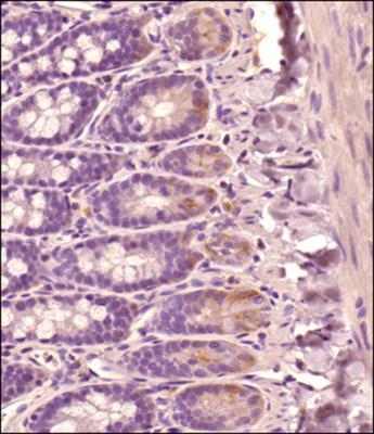
- Immunohistochemistry-Paraffin: CD44 Antibody (OX-50) [NB600-1317] - Analysis of a FFPE tissue section of Rat small intestine using Mouse anti-Rat CD44 antibody (clone OX50) at 1:100 dilution. The staining was detected using HRP-conjugated anti-Mouse secondary antibody with DAB reagent followed by hematoxylin counterstaining. This antibody clone specifically stained the membranes of the cells close to the bases of the crypts/villi which are known to actively migrate towards the tip of villi.
Supportive validation
- Submitted by
- Novus Biologicals (provider)
- Main image

- Experimental details
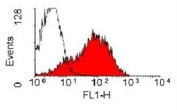
- Flow Cytometry: CD44 Antibody (OX-50) [NB600-1317] - Rat Splenocytes were stained with CD44 (OX-50) antibody NB600-1317(blue) and a matched isotype control NBP2-27287 (orange). Cells were incubated in an antibody dilution of 1 ug/mL for 20 minutes at room temperature. Both antibodies were conjugated to Alexa Fluor 488.

- Submitted by
- Novus Biologicals (provider)
- Main image
- Experimental details
- Flow Cytometry: CD44 Antibody (OX-50) [NB600-1317] - Analysis using the FITC conjugate of NB600-1317. Staining of rat spleen cells with Mouse anti Rat CD44: FITC.
