 Explore
 Validate
 Learn
LearnNBP2-29976
antibody from Novus Biologicals
Targeting: CD44
CD44R, CSPG8, HCELL, IN, MC56, MDU2, MDU3, MIC4, Pgp1
 Western blot
Antibody data
- Antibody Data
- Antigen structure
- References [0]
- Comments [0]
- Validations
- Western blot [1]
- Immunocytochemistry [1]
- Immunohistochemistry [5]
- Flow cytometry [2]
Submit
Validation data
Reference
Comment
Report error
- Product number
- NBP2-29976 - Provider product page

- Provider
- Novus Biologicals
- Product name
- Mouse Monoclonal CD44 Antibody
- Antibody type
- Monoclonal
- Description
- Protein G purified.
- Reactivity
- Human
- Host
- Mouse
- Isotype
- IgG
- Vial size
- 0.4 ml
- Concentration
- 0.71 mg/ml
- Storage
- Store at 4C short term. Aliquot and store at -20C long term. Avoid freeze-thaw cycles.
No comments: Submit comment
Supportive validation
- Submitted by
- Novus Biologicals (provider)
- Main image
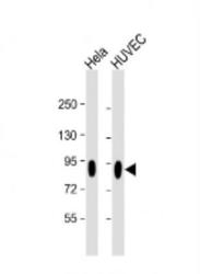
- Experimental details
- Western Blot: CD44 Antibody (Hermes-3) [NBP2-29976] - Anti-CD44 Antibody at 1:2000 dilution Lane 1: Hela whole cell lysate Lane 2: HUVEC whole cell lysate Lysates/proteins at 20 ug per lane. Secondary Goat Anti-mouse IgG, (H+L), Peroxidase conjugated at 1/10000 dilution. Predicted band size : 82 kDa Blocking/Dilution buffer: 5% NFDM/TBST.

Supportive validation
- Submitted by
- Novus Biologicals (provider)
- Main image

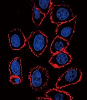
- Experimental details
- Immunocytochemistry/Immunofluorescence: CD44 Antibody (Hermes-3) [NBP2-29976] - Confocal immunofluorescent analysis with hela cell. 0.01 mg/ml primary antibody was followed by PE-conjugated goat anti-mouse lgG (whole molecule). PE emits red fluorescence. DAPI was used to stain the cell nuclear (blue).
Supportive validation
- Submitted by
- Novus Biologicals (provider)
- Main image

- Experimental details
- Immunohistochemistry: CD44 Antibody (Hermes-3) [NBP2-29976] - Immunohistochemistry analysis in formalin fixed and paraffin embedded human esophagus carcinoma followed by peroxidase conjugation of the secondary antibody and DAB staining. This data demonstrates the use of the CD44 antibody for immunohistochemistry. Clinical relevance has not been evaluated.

- Submitted by
- Novus Biologicals (provider)
- Main image
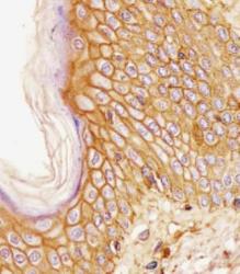
- Experimental details
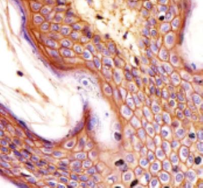
- Immunohistochemistry-Paraffin: CD44 Antibody (Hermes-3) [NBP2-29976] - Staining CD44 in human skin tissue sections by Immunohistochemistry (IHC-P - paraformaldehyde-fixed, paraffin-embedded sections). Tissue was fixed with formaldehyde and blocked with 3% BSA for 0. 5 hour at room temperature; antigen retrieval was by heat mediation with a citrate buffer (pH6). Samples were incubated with primary antibody (1/25) for 1 hours at 37 degrees C. A undiluted biotinylated goat polyvalent antibody was used as the secondary antibody.

- Submitted by
- Novus Biologicals (provider)
- Main image
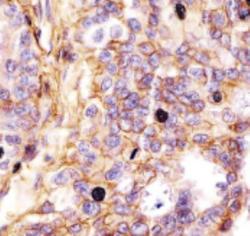
- Experimental details
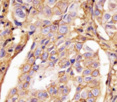
- Immunohistochemistry-Paraffin: CD44 Antibody (Hermes-3) [NBP2-29976] - Human lung adenocarcinoma tissue sections by Immunohistochemistry (IHC-P - paraformaldehyde-fixed, paraffin-embedded sections). Tissue was fixed with formaldehyde and blocked with 3% BSA for 0. 5 hour at room temperature; antigen retrieval was by heat mediation with a citrate buffer (pH6). Samples were incubated with primary antibody (1/25) for 1 hours at 37 degrees C. A undiluted biotinylated goat polyvalent antibody was used as the secondary antibody.

- Submitted by
- Novus Biologicals (provider)
- Main image

- Experimental details
- Immunohistochemistry-Paraffin: CD44 Antibody (Hermes-3) [NBP2-29976] - Human lung adenocarcinoma tissue sections by Immunohistochemistry (IHC-P - paraformaldehyde-fixed, paraffin-embedded sections). Tissue was fixed with formaldehyde and blocked with 3% BSA for 0. 5 hour at room temperature; antigen retrieval was by heat mediation with a citrate buffer (pH6). Samples were incubated with primary antibody (1/25) for 1 hours at 37 degrees C. A undiluted biotinylated goat polyvalent antibody was used as the secondary antibody.
- Submitted by
- Novus Biologicals (provider)
- Main image

- Experimental details
- Immunohistochemistry-Paraffin: CD44 Antibody (Hermes-3) [NBP2-29976] - Human skin tissue sections by Immunohistochemistry (IHC-P - paraformaldehyde-fixed, paraffin-embedded sections). Tissue was fixed with formaldehyde and blocked with 3% BSA for 0. 5 hour at room temperature; antigen retrieval was by heat mediation with a citrate buffer (pH6). Samples were incubated with primary antibody (1/25) for 1 hours at 37 degrees C. A undiluted biotinylated goat polyvalent antibody was used as the secondary antibody.
Supportive validation
- Submitted by
- Novus Biologicals (provider)
- Main image
- Experimental details
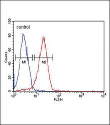
- Flow Cytometry: CD44 Antibody (Hermes-3) [NBP2-29976] - Flow cytometric analysis of Hela cells (right histogram) compared to a negative control cell (left histogram).PE-conjugated goat-anti-mouse secondary antibodies were used for the analysis.

- Submitted by
- Novus Biologicals (provider)
- Main image
- Experimental details
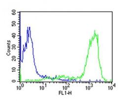
- Flow Cytometry: CD44 Antibody (Hermes-3) [NBP2-29976] - Overlay histogram showing huuman peripheral blood lymphocytes stained with CD44 antibody(green line). The cells were icubated in 2% bovine serum albumin to block non-specific protein-protein interactions followed by the antibody (1:50 dilution) for 60min at 37C. The secondary antibody used was Goat Anti-Mouse IgG, DyLight 488 Conjugated Highly Cross-Adsorbed at 1/200 dilution for 40min at 37C. Isotype control antibody (blue line) was mouse IgG2a (1ug/1x10^6 cells) used under the same conditions. Acquisition of >10, 000 events was performed.
