 Explore
 Validate
 Learn
LearnAF6127
antibody from Novus Biologicals
Targeting: CD44
CD44R, CSPG8, HCELL, IN, MC56, MDU2, MDU3, MIC4, Pgp1
 Western blot
 Immunocytochemistry
Antibody data
- Antibody Data
- Antigen structure
- References [3]
- Comments [0]
- Validations
- Western blot [2]
- Flow cytometry [4]
Submit
Validation data
Reference
Comment
Report error
- Product number
- AF6127 - Provider product page

- Provider
- Novus Biologicals
- Product name
- Sheep Polyclonal CD44 Antibody
- Antibody type
- Polyclonal
- Description
- Antigen Affinity-purified. Detects mouse and rat CD44 in direct ELISAs and Western blots. In direct ELISAs, approximately 35% cross-reactivity with recombinant human CD44 is observed.
- Reactivity
- Mouse, Rat, Porcine
- Host
- Sheep
- Conjugate
- Unconjugated
- Isotype
- IgG
- Vial size
- 100 ug
- Concentration
- LYOPH
- Storage
- Use a manual defrost freezer and avoid repeated freeze-thaw cycles. 12 months from date of receipt, -20 to -70 degreesC as supplied. 1 month, 2 to 8 degreesC under sterile conditions after reconstitution. 6 months, -20 to -70 degreesC under sterile conditions after reconstitution.
Submitted references Serglycin is involved in inflammatory response in articular mouse chondrocytes.
Deficient TSC1/TSC2-complex suppression of SOX9-osteopontin-AKT signalling cascade constrains tumour growth in tuberous sclerosis complex.
Recombinant human hyaluronidase PH20 does not stimulate an acute inflammatory response and inhibits lipopolysaccharide-induced neutrophil recruitment in the air pouch model of inflammation.
D'Ascola A, Scuruchi M, Avenoso A, Bruschetta G, Campo S, Mandraffino G, Campo GM
Biochemical and biophysical research communications 2018 May 15;499(3):506-512
Biochemical and biophysical research communications 2018 May 15;499(3):506-512
Deficient TSC1/TSC2-complex suppression of SOX9-osteopontin-AKT signalling cascade constrains tumour growth in tuberous sclerosis complex.
Jin F, Jiang K, Ji S, Wang L, Ni Z, Huang F, Li C, Chen R, Zhang H, Hu Z, Zha X
Human molecular genetics 2017 Jan 15;26(2):407-419
Human molecular genetics 2017 Jan 15;26(2):407-419
Recombinant human hyaluronidase PH20 does not stimulate an acute inflammatory response and inhibits lipopolysaccharide-induced neutrophil recruitment in the air pouch model of inflammation.
Huang Z, Zhao C, Chen Y, Cowell JA, Wei G, Kultti A, Huang L, Thompson CB, Rosengren S, Frost GI, Shepard HM
Journal of immunology (Baltimore, Md. : 1950) 2014 Jun 1;192(11):5285-95
Journal of immunology (Baltimore, Md. : 1950) 2014 Jun 1;192(11):5285-95
No comments: Submit comment
Supportive validation
- Submitted by
- Novus Biologicals (provider)
- Main image

- Experimental details
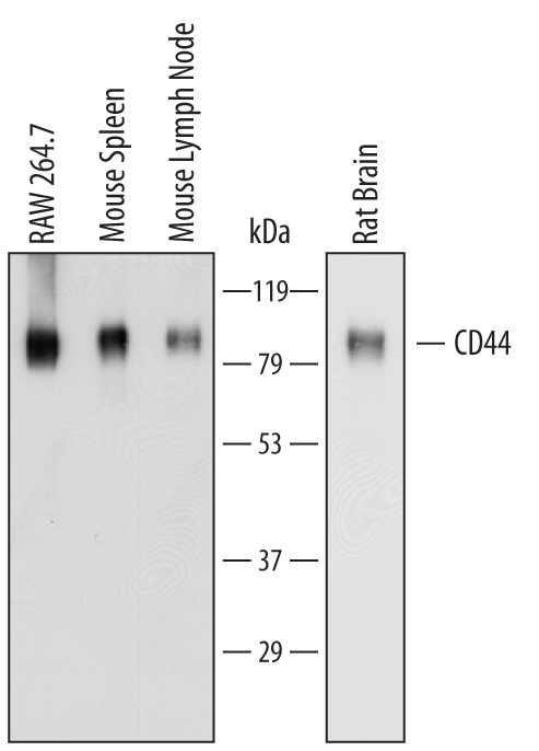
- Detection of Mouse and Rat CD44 by Western Blot. Western blot shows lysates of RAW 264.7 mouse monocyte/macrophage cell line, mouse spleen tissue, mouse lymph node tissue, and rat brain tissue. PVDF membrane was probed with 1 µg/mL of Sheep Anti-Mouse/Rat/Porcine/Equine CD44 Antigen Affinity-purified Polyclonal Antibody (Catalog # AF6127) followed by HRP-conjugated Anti-Sheep IgG Secondary Antibody (Catalog # HAF016). Specific bands were detected for CD44 at approximately 80 to 100 kDa (as indicated). This experiment was conducted under reducing conditions and using Immunoblot Buffer Group 1.
- Submitted by
- Novus Biologicals (provider)
- Main image
- Experimental details
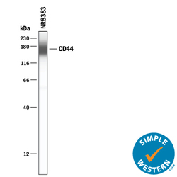
- Detection of Rat CD44 by Simple WesternTM. Simple Western lane view shows lysates of NR8383 rat alveolar macrophage cell line, loaded at 0.2 mg/mL. A specific band was detected for CD44 at approximately 169 kDa (as indicated) using 10 µg/mL of Sheep Anti-Mouse/Rat/Porcine/Equine CD44 Antigen Affinity-purified Polyclonal Antibody (Catalog # AF6127) followed by 1:50 dilution of HRP-conjugated Anti-Sheep IgG Secondary Antibody (Catalog # HAF016). This experiment was conducted under reducing conditions and using the 12-230 kDa separation system.

Supportive validation
- Submitted by
- Novus Biologicals (provider)
- Main image

- Experimental details
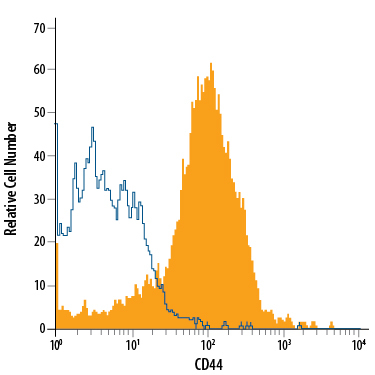
- Detection of CD44 in Equine PBMCs by Flow Cytometry. Equine peripheral blood mononuclear cells (PBMCs) were stained with Sheep Anti-Mouse/Rat/Porcine/Equine CD44 Antigen Affinity-purified Polyclonal Antibody (Catalog # AF6127, filled histogram) or isotype control antibody (Catalog # 5-001-A, open histogram), followed by Phycoerythrin-conjugated Anti-Sheep IgG Secondary Antibody (Catalog # F0126).
- Submitted by
- Novus Biologicals (provider)
- Main image

- Experimental details
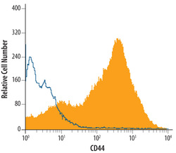
- Detection of CD44 in Porcine Mesenchymal Stem Cells by Flow Cytometry. Porcine mesenchymal stem cells were stained with Sheep Anti-Mouse/Rat/Porcine/Equine CD44 Antigen Affinity-purified Polyclonal Antibody (Catalog # AF6127, filled histogram) or isotype control antibody (Catalog # 5-001-A, open histogram), followed by Phycoerythrin-conjugated Anti-Sheep IgG Secondary Antibody (Catalog # F0126).

- Submitted by
- Novus Biologicals (provider)
- Main image

- Experimental details
- Detection of CD44 in Mouse Splenocytes by Flow Cytometry. Mouse splenocytes were stained with Sheep Anti-Mouse/Rat/Porcine/Equine CD44 Antigen Affinity-purified Polyclonal Antibody (Catalog # AF6127, filled histogram) or control antibody (Catalog # 5-001-A, open histogram), followed by Allophycocyanin-conjugated Anti-Sheep IgG Secondary Antibody (Catalog # F0127).

- Submitted by
- Novus Biologicals (provider)
- Main image
- Experimental details
- Detection of CD44 in Rat Splenocytes by Flow Cytometry. Rat splenocytes were stained with Sheep Anti-Mouse/Rat/Porcine/Equine CD44 Antigen Affinity-purified Polyclonal Antibody (Catalog # AF6127, filled histogram) or control antibody (Catalog # 5-001-A, open histogram), followed by Allophycocyanin-conjugated Anti-Sheep IgG Secondary Antibody (Catalog # F0127).
