 Explore
 Validate
 Learn
LearnV2068
antibody from NSJ Bioreagents
Targeting: CD44
CD44R, CSPG8, HCELL, IN, MC56, MDU2, MDU3, MIC4, Pgp1
 Western blot
 Flow cytometry
Antibody data
- Antibody Data
- Antigen structure
- References [0]
- Comments [0]
- Validations
- Western blot [2]
- Immunocytochemistry [1]
- Immunohistochemistry [3]
- Other assay [2]
Submit
Validation data
Reference
Comment
Report error
- Product number
- V2068 - Provider product page

- Provider
- NSJ Bioreagents
- Product name
- HCAM Antibody / CD44
- Antibody type
- Monoclonal
- Description
- This highly specific HCAM antibody is suitable for use in Flow cytometry/Immunofluorescence/Western blot/Immunohistochemistry applications with human samples.
- Reactivity
- Human
- Host
- Mouse
- Conjugate
- Unconjugated
- Antibody clone number
- 156-3C11
- Vial size
- 20 ug (with BSA and sodium azide), 100 ug (with BSA and sodium azide), 100 ug (without BSA or sodium azide), 7 ml IHC only format (if applicable)
- Concentration
- 0.2 mg/ml, 1 mg/ml
- Storage
- Store the HCAM antibody at 2-8oC (with azide) or aliquot and store at -20oC or colder (without azide).
No comments: Submit comment
Supportive validation
- Submitted by
- NSJ Bioreagents (provider)
- Main image
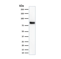
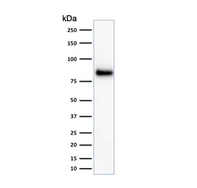
- Experimental details
- Western blot testing of HeLa cell lysate with HCAM antibody (clone 156-3C11). Predicted molecular weight ~81 kDa.
- Submitted by
- NSJ Bioreagents (provider)
- Main image
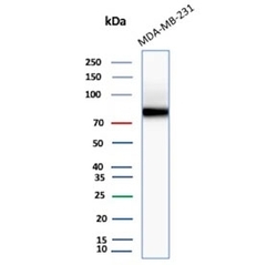
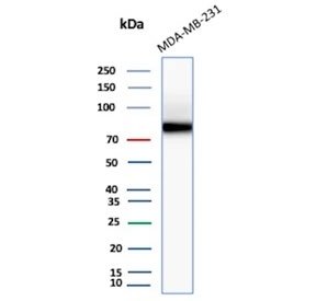
- Experimental details
- Western blot testing of MDA-MB-231 cell lysate with HCAM antibody (clone 156-3C11). Predicted molecular weight ~81 kDa.
Supportive validation
- Submitted by
- NSJ Bioreagents (provider)
- Main image
- Experimental details
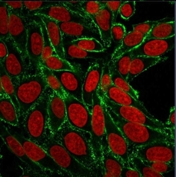
- Immunofluorescent staining of human HeLa cells with HCAM antibody (clone 156-3C11, green) and Reddot nuclear stain (red).

Supportive validation
- Submitted by
- NSJ Bioreagents (provider)
- Main image
- Experimental details
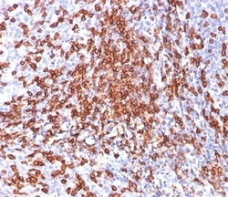
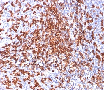
- IHC testing of FFPE human tonsil stained with HCAM antibody (clone 156-3C11).
- Submitted by
- NSJ Bioreagents (provider)
- Main image
- Experimental details
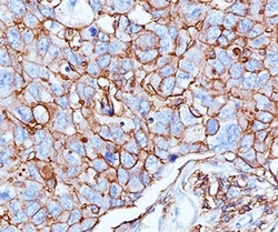
- IHC testing of human breast cancer stained with HCAM antibody (156-3C11).

- Submitted by
- NSJ Bioreagents (provider)
- Main image

- Experimental details
- IHC testing of FFPE human breast cancer stained with HCAM antibody (clone 156-3C11).

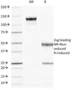
Supportive validation
- Submitted by
- NSJ Bioreagents (provider)
- Main image

- Experimental details
- SDS-PAGE Analysis of Purified, BSA-Free HCAM Antibody (clone 156-3C11). Confirmation of Integrity and Purity of the Antibody.
- Submitted by
- NSJ Bioreagents (provider)
- Main image
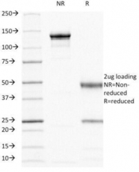
- Experimental details
- SDS-PAGE analysis of purified, BSA-free HCAM antibody (clone 156-3C11) as confirmation of integrity and purity.
