Explore
 Validate
 Learn
LearnMHCD4404
antibody from Invitrogen Antibodies
Targeting: CD44
CD44R, CSPG8, HCELL, IN, MC56, MDU2, MDU3, MIC4, Pgp1
 Flow cytometry
 Other assay
Antibody data
- Antibody Data
- Antigen structure
- References [6]
- Comments [0]
- Validations
- Other assay [1]
Submit
Validation data
Reference
Comment
Report error
- Product number
- MHCD4404 - Provider product page

- Provider
- Invitrogen Antibodies
- Product name
- CD44 Monoclonal Antibody (MEM-85), PE
- Antibody type
- Monoclonal
- Antigen
- Other
- Description
- R-phycoerythrin (PE) is a stable and highly soluble phycobiliprotein which provides maximal absorbance and fluorescence without susceptibility to internal or external fluorescence quenching, thus providing an exceptional quantum yields and molar extinction coefficients.
- Reactivity
- Human
- Host
- Mouse
- Conjugate
- Yellow dye
- Isotype
- IgG
- Antibody clone number
- MEM-85
- Vial size
- 0.5 mL
- Storage
- 4° C, store in dark
Submitted references Establishment of primary human breast cancer cell lines using "pulsed hypoxia" method and development of metastatic tumor model in immunodeficient mice.
Characterization of primary normal and malignant breast cancer cell and their response to chemotherapy and immunostimulatory agents.
Targeted introduction and effective expression of hFIX at the AAVS1 locus in mesenchymal stem cells.
Asporin Is a Fibroblast-Derived TGF-β1 Inhibitor and a Tumor Suppressor Associated with Good Prognosis in Breast Cancer.
Characterization and functionality of proliferative human Sertoli cells.
Changes in cell adhesivity and cytoskeleton-related proteins during imatinib-induced apoptosis of leukemic JURL-MK1 cells.
Nushtaeva AA, Karpushina AA, Ermakov MS, Gulyaeva LF, Gerasimov AV, Sidorov SV, Gayner TA, Yunusova AY, Tkachenko AV, Richter VA, Koval OA
Cancer cell international 2019;19:46
Cancer cell international 2019;19:46
Characterization of primary normal and malignant breast cancer cell and their response to chemotherapy and immunostimulatory agents.
Nushtaeva AA, Stepanov GA, Semenov DV, Juravlev ES, Balahonova EA, Gerasimov AV, Sidorov SV, Savelyev EI, Kuligina EV, Richter VA, Koval OA
BMC cancer 2018 Jul 9;18(1):728
BMC cancer 2018 Jul 9;18(1):728
Targeted introduction and effective expression of hFIX at the AAVS1 locus in mesenchymal stem cells.
Li SJ, Luo Y, Zhang LM, Yang W, Zhang GG
Molecular medicine reports 2017 Mar;15(3):1313-1318
Molecular medicine reports 2017 Mar;15(3):1313-1318
Asporin Is a Fibroblast-Derived TGF-β1 Inhibitor and a Tumor Suppressor Associated with Good Prognosis in Breast Cancer.
Maris P, Blomme A, Palacios AP, Costanza B, Bellahcène A, Bianchi E, Gofflot S, Drion P, Trombino GE, Di Valentin E, Cusumano PG, Maweja S, Jerusalem G, Delvenne P, Lifrange E, Castronovo V, Turtoi A
PLoS medicine 2015 Sep;12(9):e1001871
PLoS medicine 2015 Sep;12(9):e1001871
Characterization and functionality of proliferative human Sertoli cells.
Chui K, Trivedi A, Cheng CY, Cherbavaz DB, Dazin PF, Huynh AL, Mitchell JB, Rabinovich GA, Noble-Haeusslein LJ, John CM
Cell transplantation 2011;20(5):619-35
Cell transplantation 2011;20(5):619-35
Changes in cell adhesivity and cytoskeleton-related proteins during imatinib-induced apoptosis of leukemic JURL-MK1 cells.
Kuželová K, Pluskalová M, Grebeňová D, Pavlásková K, Halada P, Hrkal Z
Journal of cellular biochemistry 2010 Dec 15;111(6):1413-25
Journal of cellular biochemistry 2010 Dec 15;111(6):1413-25
No comments: Submit comment
Supportive validation
- Submitted by
- Invitrogen Antibodies (provider)
- Main image

- Experimental details
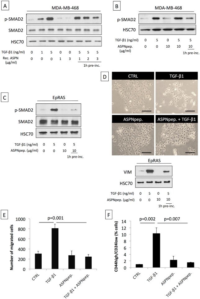
- Fig 4 Asporin binds to TGF-beta1 and inhibits its downstream signaling and function. (A) Western blot analysis of phospho-SMAD2 (p-SMAD2) and SMAD2 total protein extracts from MDA-MB-468 breast cancer cells treated for 15 min with TGF-beta1 and/or human recombinant asporin (Rec. ASPN). (B) Western blot analysis of p-SMAD2 in total protein extracts from MDA-MB-468 breast cancer cells treated with TGF-beta1 and/or asporin peptide corresponding to the 159-205 amino acid region (ASPNpep.). (C) Western blot analysis of p-SMAD2 and SMAD2 in total protein extracts from EpRAS cells treated for 15 min with TGF-beta1 (5 ng/ml) and/or asporin peptide. (D) EMT induction in EpRAS cells in the presence of TGF-beta1 and/or asporin peptide. EMT was monitored both at the phenotype level (upper panel) and using Western blot evaluation of VIM expression in total protein extracts from EpRAS cells (lower panel). (A-D): HSC70 was used as loading control. (E) Transwell migration assay of EpRAS cells pretreated with TGF-beta1 (5 ng/ml) and/or asporin peptide (10 mug/ml). (F) Quantification of the CSC population in EpRAS cells following TGF-beta1 and/or asporin peptide treatment. (E and F): The data are presented as mean +- SD. All panels: statistical significance was calculated using the Student's t -test (as described in the Methods section). Western blots show representative data of three independent experiments.
- Conjugate
- Yellow dye