Explore
 Validate
 Learn
LearnMA5-15462
antibody from Invitrogen Antibodies
Targeting: CD44
CD44R, CSPG8, HCELL, IN, MC56, MDU2, MDU3, MIC4, Pgp1
 Western blot
 ELISA
ELISA Immunocytochemistry
Antibody data
- Antibody Data
- Antigen structure
- References [2]
- Comments [0]
- Validations
- Immunocytochemistry [4]
- Immunohistochemistry [5]
- Flow cytometry [2]
- Other assay [2]
Submit
Validation data
Reference
Comment
Report error
- Product number
- MA5-15462 - Provider product page

- Provider
- Invitrogen Antibodies
- Product name
- CD44 Monoclonal Antibody (8E2F3)
- Antibody type
- Monoclonal
- Antigen
- Purifed from natural sources
- Description
- MA5-15462 targets CD44 in indirect ELISA, FACS, IF, IHC, and WB applications and shows reactivity with Human and mouse samples. The MA5-15462 immunogen is purified recombinant fragment of human CD44 (628-699) expressed in E. Coli. MA5-15462 detects CD44 which has a predicted molecular weight of approximately 82kDa.
- Reactivity
- Human, Mouse
- Host
- Mouse
- Isotype
- IgG
- Antibody clone number
- 8E2F3
- Vial size
- 100 μL
- Concentration
- Conc. Not Determined
- Storage
- Store at 4°C short term. For long term storage, store at -20°C, avoiding freeze/thaw cycles.
Submitted references Insights into homeobox B9: a propeller for metastasis in dormant prostate cancer progenitor cells.
Heterogeneity and dynamics of active Kras-induced dysplastic lineages from mouse corpus stomach.
Sui Y, Hu W, Zhang W, Li D, Zhu H, You Q, Zhu R, Yi Q, Tang T, Gao L, Zhu S, Yang T
British journal of cancer 2021 Sep;125(7):1003-1015
British journal of cancer 2021 Sep;125(7):1003-1015
Heterogeneity and dynamics of active Kras-induced dysplastic lineages from mouse corpus stomach.
Min J, Vega PN, Engevik AC, Williams JA, Yang Q, Patterson LM, Simmons AJ, Bliton RJ, Betts JW, Lau KS, Magness ST, Goldenring JR, Choi E
Nature communications 2019 Dec 5;10(1):5549
Nature communications 2019 Dec 5;10(1):5549
No comments: Submit comment
Supportive validation
- Submitted by
- Invitrogen Antibodies (provider)
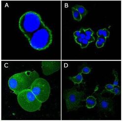
- Main image
- Experimental details
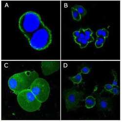
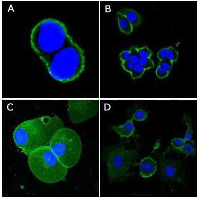
- Immunofluorescence analysis of methanol-fixed A431 (A), HeLa (B), PANC-1 (C) and EC (D) cells using CD44/H-CAM monoclonal antibody (Product # MA5-15462) (Green). Blue: DRAQ5 fluorescent DNA dye.
- Submitted by
- Invitrogen Antibodies (provider)
- Main image

- Experimental details
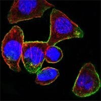
- Immunofluorescence analysis of PANC-1 cells using CD44/H-CAM monoclonal antibody (Product # MA5-15462) (Green). Blue: DRAQ5 fluorescent DNA dye. Red: Actin filaments have been labeled with DY-554 phalloidin.
- Submitted by
- Invitrogen Antibodies (provider)
- Main image

- Experimental details
- Immunofluorescence analysis of PANC-1 cells using CD44/H-CAM monoclonal antibody (Product # MA5-15462) (Green). Blue: DRAQ5 fluorescent DNA dye. Red: Actin filaments have been labeled with DY-554 phalloidin.

- Submitted by
- Invitrogen Antibodies (provider)
- Main image
- Experimental details
- Immunofluorescence analysis of methanol-fixed A431 (A), HeLa (B), PANC-1 (C) and EC (D) cells using CD44/H-CAM monoclonal antibody (Product # MA5-15462) (Green). Blue: DRAQ5 fluorescent DNA dye.

Supportive validation
- Submitted by
- Invitrogen Antibodies (provider)
- Main image
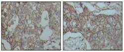
- Experimental details
- Immunohistochemical analysis of paraffin-embedded human breast carcinoma tissues using CD44/H-CAM monoclonal antibody (Product # MA5-15462) followed with DAB staining
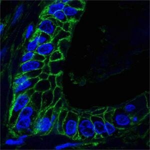
- Submitted by
- Invitrogen Antibodies (provider)
- Main image

- Experimental details
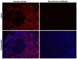
- Immunofluorescene analysis of paraffin-embedded human lung cancer tissues using CD44/H-CAM monoclonal antibody (Product # MA5-15462) (Green). Blue: DRAQ5 fluorescent DNA dye.
- Submitted by
- Invitrogen Antibodies (provider)
- Main image
- Experimental details
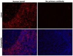
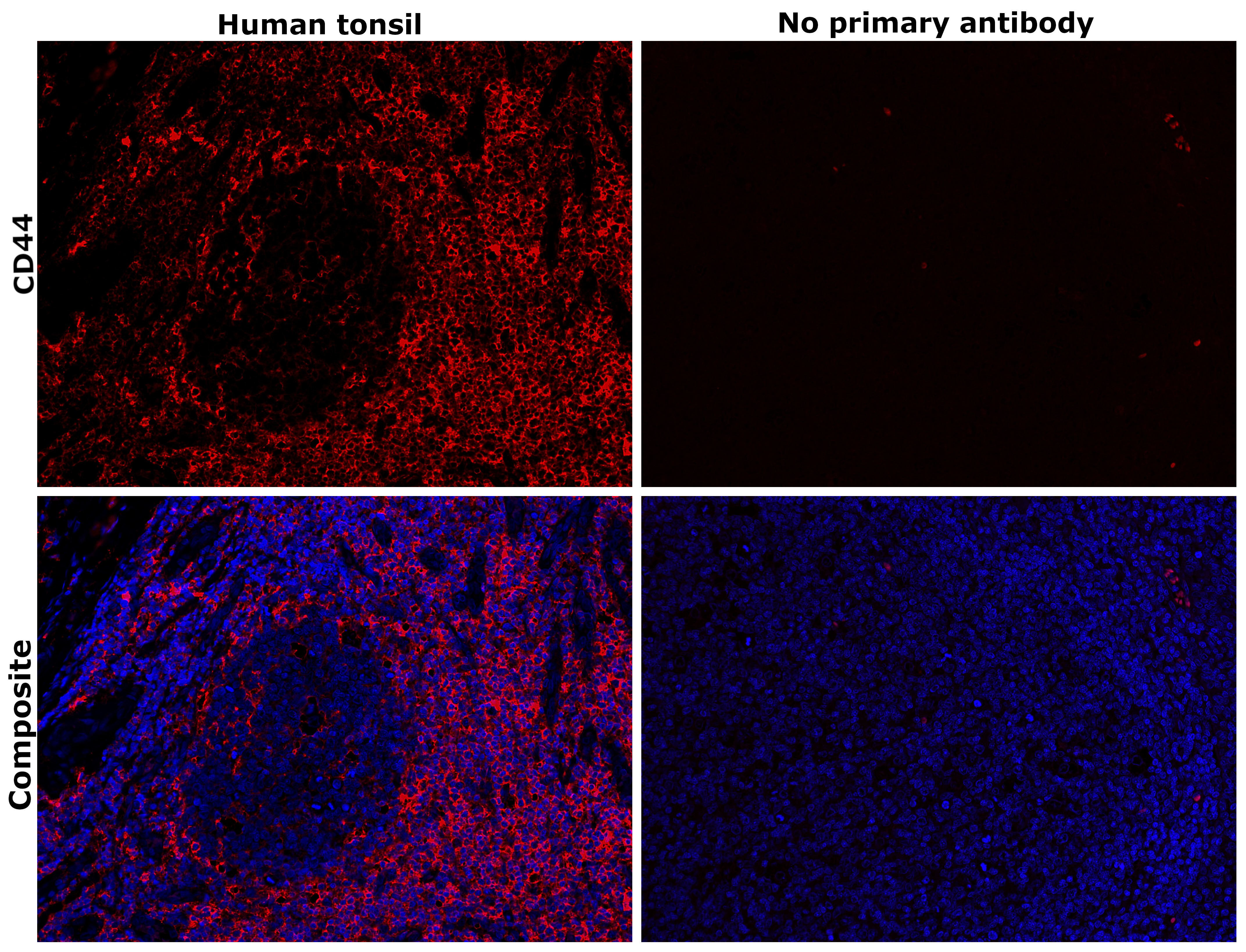
- Immunohistochemical analysis of CD44 was performed using formalin-fixed paraffin-embedded human tonsil tissue sections. To expose the target protein, heat-induced epitope retrieval was performed on de-paraffinized sections using eBioscience™ IHC Antigen Retrieval Solution - High pH (10X) (Product # 00-4956-58) diluted to 1X solution in water in a decloaking chamber at 110 degree Celsius for 15 minutes. Following antigen retrieval, the sections were blocked with 3% H2O2 for 1 hour at room temperature followed by 2% normal goat serum in 1X PBS for 45 minutes at room temperature and then probed with or without CD44 Monoclonal Antibody (8E2F3) (Product # MA5-15462) at 1:500 dilution in 0.1% normal goat serum overnight at 4 degree Celsius in a humidified chamber. Detection was performed using Alexa Fluor™ 647 Tyramide SuperBoost™ Kit, goat anti-mouse IgG (Product # B40916). Nuclei were stained with DAPI (Product # D1306) and the sections were mounted using ProLong™ Glass Antifade Mountant (Product # P36984). The images were captured on EVOS™ M7000 Imaging System (Product # AMF7000) at 20X magnification and externally deconvoluted.
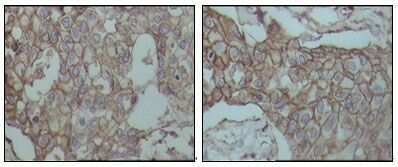
- Submitted by
- Invitrogen Antibodies (provider)
- Main image
- Experimental details
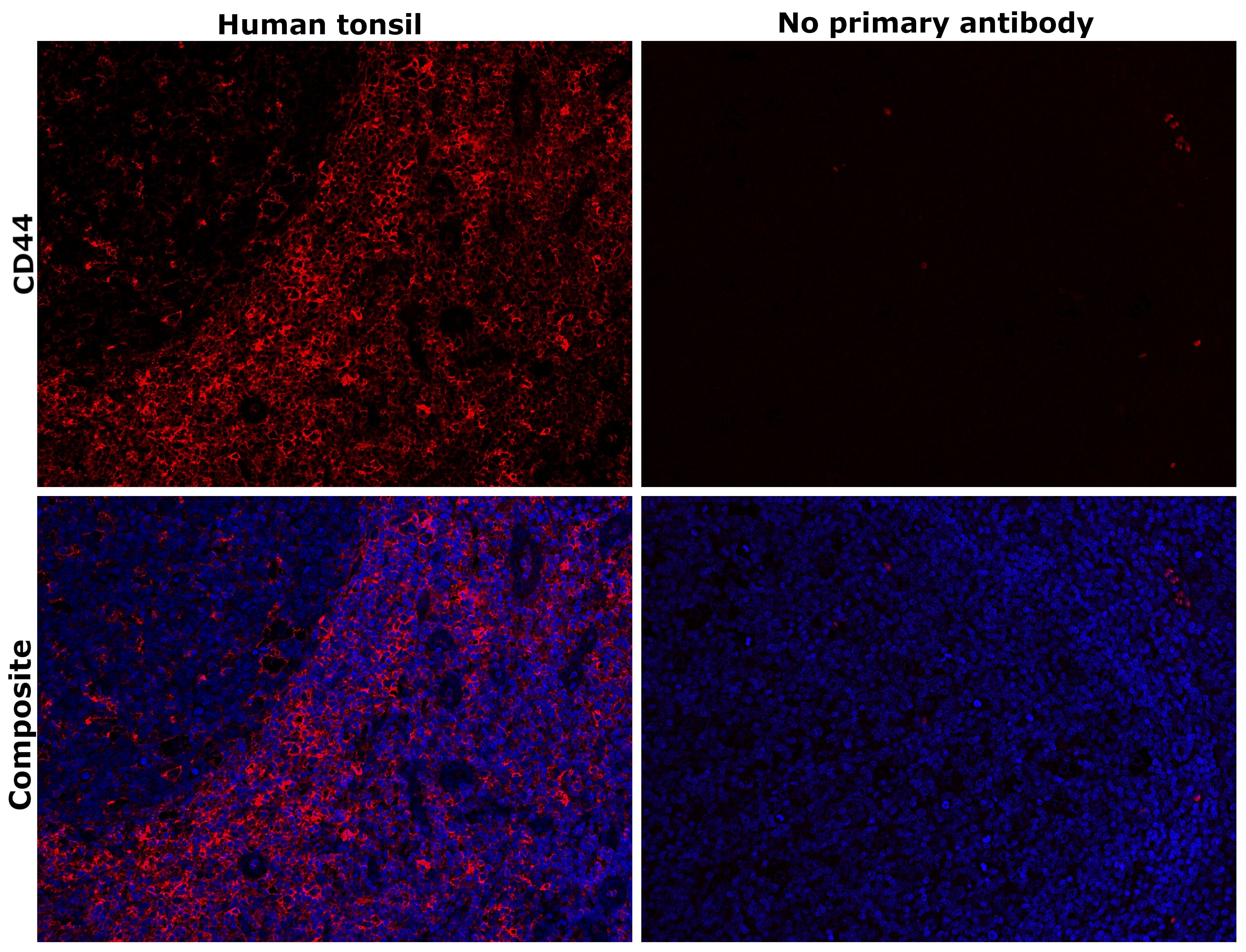
- Immunohistochemical analysis of CD44 was performed using formalin-fixed paraffin-embedded human tonsil tissue sections. To expose the target protein, heat-induced epitope retrieval was performed on de-paraffinized sections using eBioscience™ IHC Antigen Retrieval Solution - Low pH (10X) (Product # 00-4955-58) diluted to 1X solution in water in a decloaking chamber at 110 degree Celsius for 15 minutes. Following antigen retrieval, the sections were blocked with 3% H2O2 for 1 hour at room temperature followed by 2% normal goat serum in 1X PBS for 45 minutes at room temperature and then probed with or without CD44 Monoclonal Antibody (8E2F3) (Product # MA5-15462) at 1:500 dilution in 0.1% normal goat serum overnight at 4 degree Celsius in a humidified chamber. Detection was performed using Alexa Fluor™ 647 Tyramide SuperBoost™ Kit, goat anti-mouse IgG (Product # B40916). Nuclei were stained with DAPI (Product # D1306) and the sections were mounted using ProLong™ Glass Antifade Mountant (Product # P36984). The images were captured on EVOS™ M7000 Imaging System (Product # AMF7000) at 20X magnification and externally deconvoluted.

- Submitted by
- Invitrogen Antibodies (provider)
- Main image
- Experimental details
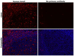
- Immunohistochemical analysis of CD44 was performed using formalin-fixed paraffin-embedded human tonsil tissue sections. To expose the target protein, heat-induced epitope retrieval was performed on de-paraffinized sections using eBioscience™ IHC Antigen Retrieval Solution - High pH (10X) (Product # 00-4956-58) diluted to 1X solution in water in a decloaking chamber at 110 degree Celsius for 15 minutes. Following antigen retrieval, the sections were blocked with 3% H2O2 for 1 hour at room temperature followed by 2% normal goat serum in 1X PBS for 45 minutes at room temperature and then probed with or without CD44 Monoclonal Antibody (8E2F3) (Product # MA5-15462) at 1:500 dilution in 0.1% normal goat serum overnight at 4 degree Celsius in a humidified chamber. Detection was performed using Alexa Fluor™ 647 Tyramide SuperBoost™ Kit, goat anti-mouse IgG (Product # B40916). Nuclei were stained with DAPI (Product # D1306) and the sections were mounted using ProLong™ Glass Antifade Mountant (Product # P36984). The images were captured on EVOS™ M7000 Imaging System (Product # AMF7000) at 20X magnification and externally deconvoluted.
Supportive validation
- Submitted by
- Invitrogen Antibodies (provider)
- Main image

- Experimental details
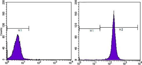
- Flow cytometric analysis of HeLa cells using CD44/H-CAM monoclonal antibody (Product # MA5-15462) (right) and negative control (left).
- Submitted by
- Invitrogen Antibodies (provider)
- Main image
- Experimental details
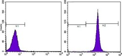
- Flow cytometric analysis of HeLa cells using CD44/H-CAM monoclonal antibody (Product # MA5-15462) (right) and negative control (left).

Supportive validation
- Submitted by
- Invitrogen Antibodies (provider)
- Main image
- Experimental details
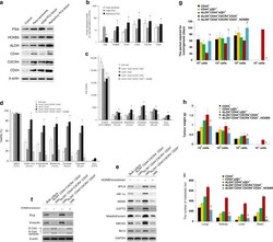
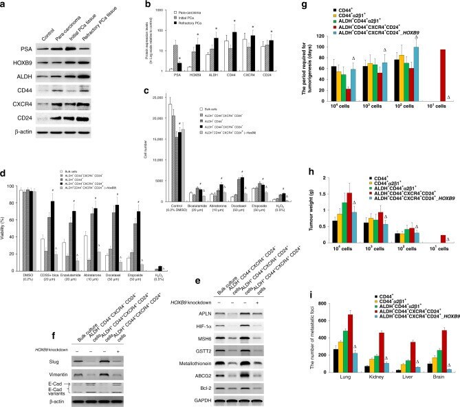
- Fig. 6 The ALDH + CD44 + CXCR4 + CD24 + subpopulation in human PCa tissues. a Western blot analysis was performed to determine the protein expressions of PSA, HOXB9, ALDH, CD44, CXCR4 and CD24 in the controls (PCa tissue with Gleason score 6), para-carcinoma (2 mm away from PCa tissue), initial PCa tissue (derived from PCa at first diagnosis via radical prostatectomy) and refractory PCa tissue (derived after recurrence), respectively. beta-actin was used as an internal control. b Quantification of ( a ). * P < 0.05 vs. initial PCa tissues. ( n = 6). c Human PCa tissue was subcutaneously implanted into NOD-SCID mice to establish a patient-derived xenograft (PDX) model. Subsets of cells (as indicated) were derived from the PDX model and seeded in 96-well plates (1 x 10 4 cells/well) and treated with different anti-androgens (as indicated) and chemotherapeutic agents, with 0.2% DMSO and 0.5% H 2 O 2 were used as negative and positive controls, respectively. After 48 h of treatment, cells were incubated with alamarBlue solution for 4 h, and cell viability was measured with excitation wavelength at 530-560 nm and emission wavelength at 590 nm using a TECAN Infinite 200 PRO microplate reader. d Subsets of cells (as indicated) were derived from the PDX model and seeded in 96-well plates (1 x 10 4 cells/well). Cells were treated with different chemotherapeutic drugs, as indicated, for 72 h. Then, a WST-1 proliferation assay was performed. The absorbance was measured at 450 nm using a
- Submitted by
- Invitrogen Antibodies (provider)
- Main image
- Experimental details
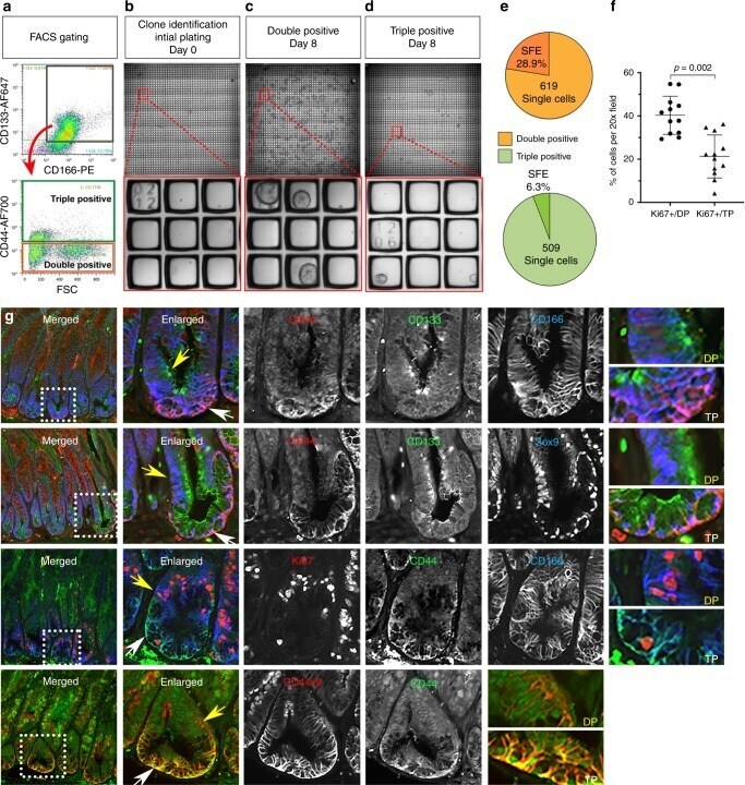
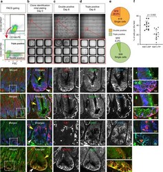
- Fig. 8 Identification of dysplastic stem cells in Meta4. a - e Clonal sphere forming efficiency from Meta4 subpopulations. a Meta4 organoids were dissociated to single cells, stained for CD44, CD133, CD166, and FACS isolated. Following doublet-discrimination and dead cell exclusion, CD133/CD166 positive cells (gray gate) were applied (red arrow) to a CD44 positive gate to distinguish between double-positive CD44neg/CD133+/CD166+ (orange gate) and triple-positive CD44+/CD133+/CD166+cells (green gate). Double-positive and Triple-positive cells were collected by FACS and applied to CRAs. b Immediately after plating, single cells were quantified and their physical address on the CRA was determined. c , d After 8 days of culture, spheres were quantified and their physical address on the CRA was determined. Upper-panels are low magnification of each CRA containing 2025 wells. Red-boxes indicate the regions in the higher magnification images depicted in the lower panels. e Spheres-Forming Efficiency (SFE) of clonal events was determined by quantifying the number of spheres that developed from all single cell events at t = 0. There were 619 Double-positive clonal events identified in the CRAs, and 28.9% of those generated spheres (orange chart). There were 509 Triple-positive clonal events identified in the CRAs, and 6.3% of those generated spheres (green chart). The data represent binary outcomes. f Quantitation of immunofluorescence staining of Ki67-positive cells in either Double-