Explore
 Validate
 Learn
Learn Western blot
Antibody data
- Antibody Data
- Antigen structure
- References [0]
- Comments [0]
- Validations
- Western blot [2]
- Immunocytochemistry [1]
- Immunohistochemistry [1]
Submit
Validation data
Reference
Comment
Report error
- Product number
- PA5-79522 - Provider product page

- Provider
- Invitrogen Antibodies
- Product name
- ISG15 Polyclonal Antibody
- Antibody type
- Polyclonal
- Antigen
- Recombinant full-length protein
- Description
- Reconstitute with 0.2 mL of distilled water to yield a concentration of 500 µg/mL.
- Reactivity
- Human
- Host
- Rabbit
- Isotype
- IgG
- Vial size
- 100 µg
- Concentration
- 500 µg/mL
- Storage
- -20°C
No comments: Submit comment
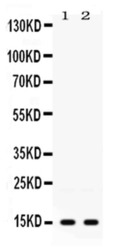
Supportive validation
- Submitted by
- Invitrogen Antibodies (provider)
- Main image
- Experimental details
- Western blot analysis of ISG15 in 22RV1 whole cell lysate (lane 1) and HeLa whole cell lysate (lane 2). Sample was incubated with ISG15 polyclonal antibody (Product # PA5-79522) at a dilution of 0.5 µg/mL. Signal development was performed using a chemiluminescence (ECL) kit.

- Submitted by
- Invitrogen Antibodies (provider)
- Main image
- Experimental details
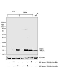
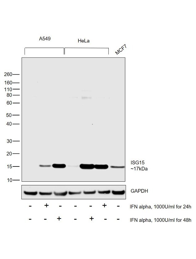
- Western blot was performed using Anti-ISG15 Rabbit Polyclonal Antibody (Product # PA5-79522) and a 17kDa band corresponding to ISG15 was observed across cell lines tested and increased upon IFN alpha treatment. Whole cell extracts (30 µg lysate) of A549 (Lane 1), A549 treated with IFN alpha (1000U/ml for 24h) (Lane 2), A549 treated with IFN alpha (1000U/ml for 48h) (Lane 3), HeLa (Lane 4), HeLa treated with IFN alpha (1000U/ml for 48h) (Lane 5), HeLa treated with IFN alpha (1000U/ml for 24h (Lane 6) and MCF7 (Lane 7) were electrophoresed using Novex® NuPAGE® 4-12 % Bis-Tris gel (Product # NP0322BOX). Resolved proteins were then transferred onto a nitrocellulose membrane (Product # IB23001) by iBlot® 2 Dry Blotting System (Product # IB21001). The blot was probed with the primary antibody (0.25ug/ml) and detected by chemiluminescence with Goat anti-Rabbit IgG (H+L), Superclonal™ Recombinant Secondary Antibody, HRP (Product # A27036, 1:4000 dilution) using the iBright FL 1000 (Product # A32752). Chemiluminescent detection was performed using Novex® ECL Chemiluminescent Substrate Reagent Kit (Product # WP20005).
Supportive validation
- Submitted by
- Invitrogen Antibodies (provider)
- Main image

- Experimental details
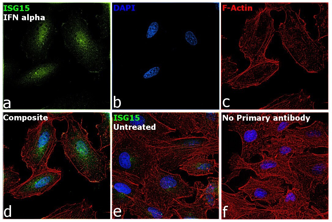
- Immunofluorescence analysis of ISG15 was performed using 70% confluent log phase A549 cells treated with IFN alpha (1000U/mL for 48h). The cells were fixed with 4% paraformaldehyde for 10 minutes, permeabilized with 0.1% Triton™ X-100 for 15 minutes, and blocked with 1% BSA for 1 hour at room temperature. The cells were labeled with ISG15 Rabbit Polyclonal Antibody (Product # PA5-79522) at 1:100 dilution in 0.1% BSA, incubated at 4 degree Celsius overnight and then labeled with Goat anti-Rabbit IgG (H+L) Superclonal™ Recombinant Secondary Antibody, Alexa Fluor® 488 conjugate (Product # A27034) at a dilution of 1:2000 for 45 minutes at room temperature (Panel a: green). Nuclei (Panel b: blue) were stained with SlowFade® Gold Antifade Mountant with DAPI (Product # S36938). F-actin (Panel c: red) was stained with Rhodamine Phalloidin (Product # R415, 1:300). Panel d represents the merged image showing increased ISG15 expression and localization to nucleus and cytoplasm. Panel e shows untreated cells with lower expression of ISG15. Panel f represents control cells with no primary antibody to assess background. The images were captured at 60X magnification.
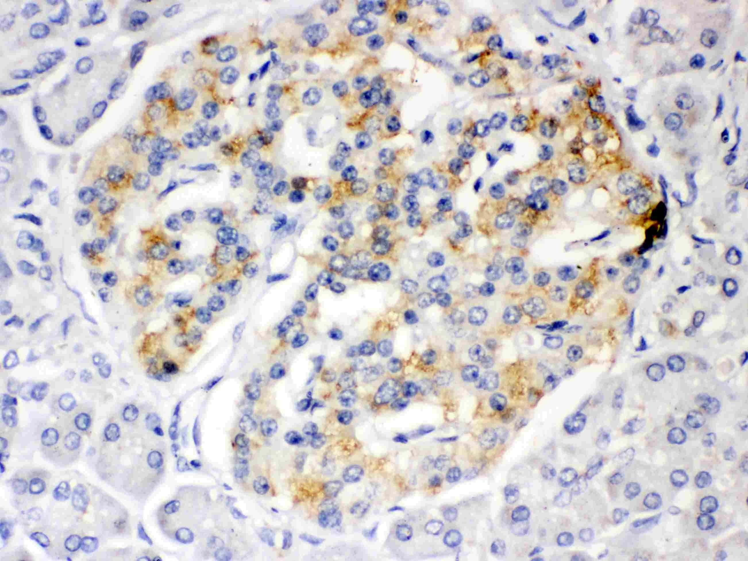
Supportive validation
- Submitted by
- Invitrogen Antibodies (provider)
- Main image
- Experimental details
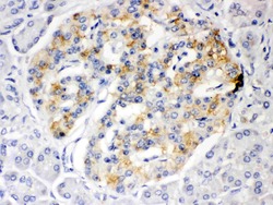
- Immunohistochemistry analysis of ISG15 on paraffin-embedded human pancreatic cancer tissue. Sample was incubated with ISG15 polyclonal antibody (Product# PA5-79522) with a dilution of 1 µg/mL, and developed by Streptavidin-Biotin-Complex (SABC) with DAB chromogen method.