 Explore
 Validate
 Learn
Learn Western blot
Antibody data
- Antibody Data
- Antigen structure
- References [0]
- Comments [0]
- Validations
- Western blot [1]
- Immunocytochemistry [1]
- Immunohistochemistry [2]
Submit
Validation data
Reference
Comment
Report error
- Product number
- PA5-22969 - Provider product page

- Provider
- Invitrogen Antibodies
- Product name
- GPX3 Polyclonal Antibody
- Antibody type
- Polyclonal
- Antigen
- Other
- Description
- The target sequence has 89% sequence homology with mouse, 88% sequence homology with bovine and 84% sequence homology with porcine. Suggested positive control: HeLa whole cell extract.
- Reactivity
- Human, Rat
- Host
- Rabbit
- Isotype
- IgG
- Vial size
- 100 μL
- Concentration
- Conc. Not Determined
- Storage
- Store at 4°C short term. For long term storage, store at -20°C, avoiding freeze/thaw cycles.
No comments: Submit comment
Supportive validation
- Submitted by
- Invitrogen Antibodies (provider)
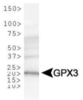
- Main image
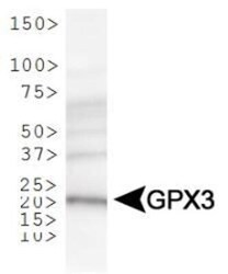
- Experimental details
- Western blot analysis of GPX3 in human kidney lysate. Sample was incubated in GPX3 polyclonal antibody (Product # PA5-22969).
Supportive validation
- Submitted by
- Invitrogen Antibodies (provider)
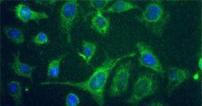
- Main image
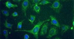
- Experimental details
- Immunofluorescent analysis of GPX3 using a polyclonal antibody (Product # PA5-22969).
Supportive validation
- Submitted by
- Invitrogen Antibodies (provider)
- Main image

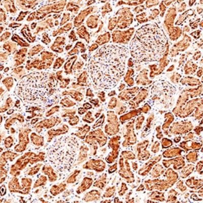
- Experimental details
- Immunohistochemical analysis of GPX3 in formalin fixed paraffin-embedded (FFPE) human kidney. Samples were incubated in GPX3 polyclonal antibody (Product # PA5-22969) using a dilution of 1:200. Bond Rx autostainer (Leica Biosystems). The assay involved 20 minutes of heat induced antigen retrieval (HIER) using 10mM sodium citrate buffer (pH 6.0) and endogenous peroxidase quenching with peroxide block. The sections were incubated with primary antibody for 30 minutes and Bond Polymer Refine Detection (Leica Biosystems) with DAB was used for signal development followed by counterstaining with hematoxylin. Whole slide scanning and capturing of representative images was performed using Aperio AT2 (Leica Biosystems). Cytoplasmic staining of Glutathione Peroxidase 3 was observed. Staining was performed by Histowiz.
- Submitted by
- Invitrogen Antibodies (provider)
- Main image
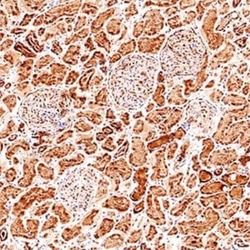
- Experimental details
- Immunohistochemical analysis of GPX3 in formalin fixed paraffin-embedded (FFPE) human kidney. Samples were incubated in GPX3 polyclonal antibody (Product # PA5-22969) using a dilution of 1:200. Bond Rx autostainer (Leica Biosystems). The assay involved 20 minutes of heat induced antigen retrieval (HIER) using 10mM sodium citrate buffer (pH 6.0) and endogenous peroxidase quenching with peroxide block. The sections were incubated with primary antibody for 30 minutes and Bond Polymer Refine Detection (Leica Biosystems) with DAB was used for signal development followed by counterstaining with hematoxylin. Whole slide scanning and capturing of representative images was performed using Aperio AT2 (Leica Biosystems). Cytoplasmic staining of Glutathione Peroxidase 3 was observed. Staining was performed by Histowiz.
