Explore
 Validate
 Learn
Learn Western blot
Antibody data
- Antibody Data
- Antigen structure
- References [3]
- Comments [0]
- Validations
- Western blot [2]
- Immunohistochemistry [1]
Submit
Validation data
Reference
Comment
Report error
- Product number
- AF4199 - Provider product page

- Provider
- R&D Systems
- Product name
- Human/Mouse/Rat Glutathione Peroxidase 3/GPX3 Antibody
- Antibody type
- Polyclonal
- Description
- Antigen Affinity-purified. Detects human, mouse, and rat Glutathione Peroxidase 3/GPX3 in Western blots and detects recombinant human and recombinant mouse Glutathione Peroxidase 3/GPX3 in direct ELISAs. In direct ELISAs, approximately 15% cross-reactivity with recombinant human (rh) GPX5 and recombinant mouse (rm) GPX1 is observed, and less than 4% cross-reactivity with rhGPX4, rhGPX6, and rmGPX2 is observed.
- Reactivity
- Human, Mouse, Rat
- Host
- Goat
- Conjugate
- Unconjugated
- Antigen sequence
P46412- Isotype
- IgG
- Vial size
- 100 ug
- Concentration
- LYOPH
- Storage
- Use a manual defrost freezer and avoid repeated freeze-thaw cycles. 12 months from date of receipt, -20 to -70 °C as supplied. 1 month, 2 to 8 °C under sterile conditions after reconstitution. 6 months, -20 to -70 °C under sterile conditions after reconstitution.
Submitted references Transcript and protein analysis reveals better survival skills of monocyte-derived dendritic cells compared to monocytes during oxidative stress.
Disruption of the selenocysteine lyase-mediated selenium recycling pathway leads to metabolic syndrome in mice.
A role for GPx3 in activity of normal and leukemia stem cells.
Van Brussel I, Schrijvers DM, Martinet W, Pintelon I, Deschacht M, Schnorbusch K, Maes L, Bosmans JM, Vrints CJ, Adriaensen D, Cos P, Bult H
PloS one 2012;7(8):e43357
PloS one 2012;7(8):e43357
Disruption of the selenocysteine lyase-mediated selenium recycling pathway leads to metabolic syndrome in mice.
Seale LA, Hashimoto AC, Kurokawa S, Gilman CL, Seyedali A, Bellinger FP, Raman AV, Berry MJ
Molecular and cellular biology 2012 Oct;32(20):4141-54
Molecular and cellular biology 2012 Oct;32(20):4141-54
A role for GPx3 in activity of normal and leukemia stem cells.
Herault O, Hope KJ, Deneault E, Mayotte N, Chagraoui J, Wilhelm BT, Cellot S, Sauvageau M, Andrade-Navarro MA, Hébert J, Sauvageau G
The Journal of experimental medicine 2012 May 7;209(5):895-901
The Journal of experimental medicine 2012 May 7;209(5):895-901
No comments: Submit comment
Supportive validation
- Submitted by
- R&D Systems (provider)
- Main image
- Experimental details
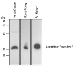
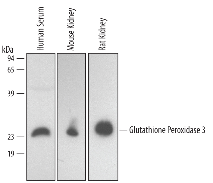
- Detection of Human/Mouse/Rat Glutathione Peroxidase 3 by Western Blot. Western blot shows lysates of human plasma, mouse kidney tissue, and rat kidney tissue. PVDF membrane was probed with 0.5 µg/mL of Goat Anti-Human/Mouse/Rat Glutathione Peroxidase 3 Antigen Affinity-purified Polyclonal Antibody (Catalog # AF4199) followed by HRP-conjugated Anti-Goat IgG Secondary Antibody (Catalog # HAF017). A specific band was detected for Glutathione Peroxidase 3 at approximately 24kDa (as indicated). This experiment was conducted under reducing conditions and using Immunoblot Buffer Group 1.
- Submitted by
- R&D Systems (provider)
- Main image
- Experimental details
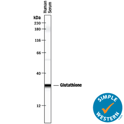
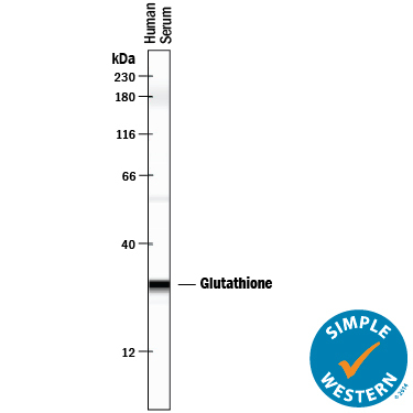
- Detection of Human Glutathione Peroxidase 3/GPX3 by Simple WesternTM. Simple Western lane view shows human serum, loaded at 0.2 mg/mL. A specific band was detected for Glutathione Peroxidase 3/GPX3 at approximately 29 kDa (as indicated) using 25 µg/mL of Goat Anti-Human/Mouse/Rat Glutathione Peroxidase 3/GPX3 Antigen Affinity-purified Polyclonal Antibody (Catalog # AF4199) followed by 1:50 dilution of HRP-conjugated Anti-Goat IgG Secondary Antibody (Catalog # HAF109). This experiment was conducted under reducing conditions and using the 12-230 kDa separation system.
Supportive validation
- Submitted by
- R&D Systems (provider)
- Main image
- Experimental details
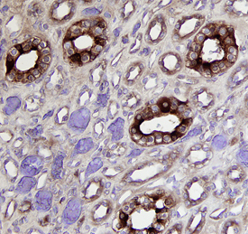
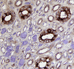
- Glutathione Peroxidase 3/GPX3 in Human Kidney. Glutathione Peroxidase 3/GPX3 was detected in immersion fixed paraffin-embedded sections of human kidney using 15 µg/mL Goat Anti-Human/Mouse/Rat Glutathione Peroxidase 3/GPX3 Antigen Affinity-purified Polyclonal Antibody (Catalog # AF4199) overnight at 4 °C. Tissue was stained with the Anti-Goat HRP-DAB Cell & Tissue Staining Kit (brown; Catalog # CTS008) and counterstained with hematoxylin (blue). Specific labeling was localized to the cytoplasm in tubules. View our protocol for Chromogenic IHC Staining of Paraffin-embedded Tissue Sections.