Explore
 Validate
 Learn
Learn Western blot
 Immunocytochemistry
Antibody data
- Antibody Data
- Antigen structure
- References [4]
- Comments [0]
- Validations
- Immunocytochemistry [1]
Submit
Validation data
Reference
Comment
Report error
- Product number
- HPA001247 - Provider product page

- Provider
- Atlas Antibodies
- Proper citation
- Atlas Antibodies Cat#HPA001247, RRID:AB_1078602
- Product name
- Anti-CYC1
- Antibody type
- Polyclonal
- Description
- Polyclonal Antibody against Human CYC1, Gene description: cytochrome c-1, Alternative Gene Names: UQCR4, Validated applications: WB, ICC, IHC, Uniprot ID: P08574, Storage: Store at +4°C for short term storage. Long time storage is recommended at -20°C.
- Reactivity
- Human, Mouse, Rat
- Host
- Rabbit
- Conjugate
- Unconjugated
- Isotype
- IgG
- Vial size
- 100 µl
- Concentration
- 0.1 mg/ml
- Storage
- Store at +4°C for short term storage. Long time storage is recommended at -20°C.
- Handling
- The antibody solution should be gently mixed before use.
Submitted references C11orf83, a Mitochondrial Cardiolipin-Binding Protein Involved in bc1 Complex Assembly and Supercomplex Stabilization
Applying Sodium Carbonate Extraction Mass Spectrometry to Investigate Defects in the Mitochondrial Respiratory Chain
Effects of moderate global maternal nutrient reduction on fetal baboon renal mitochondrial gene expression at 0.9 gestation
Variance decomposition of protein profiles from antibody arrays using a longitudinal twin model
Desmurs M, Foti M, Raemy E, Vaz F, Martinou J, Bairoch A, Lane L
Molecular and Cellular Biology 2023;35(7):1139-1156
Molecular and Cellular Biology 2023;35(7):1139-1156
Applying Sodium Carbonate Extraction Mass Spectrometry to Investigate Defects in the Mitochondrial Respiratory Chain
Robinson D, Hock D, Muellner-Wong L, Kugapreethan R, Reljic B, Surgenor E, Rodrigues C, Caruana N, Stroud D
Frontiers in Cell and Developmental Biology 2022;10
Frontiers in Cell and Developmental Biology 2022;10
Effects of moderate global maternal nutrient reduction on fetal baboon renal mitochondrial gene expression at 0.9 gestation
Pereira S, Oliveira P, Tavares L, Moreno A, Cox L, Nathanielsz P, Nijland M
American Journal of Physiology-Renal Physiology 2015;308(11):F1217-F1228
American Journal of Physiology-Renal Physiology 2015;308(11):F1217-F1228
Variance decomposition of protein profiles from antibody arrays using a longitudinal twin model
Kato B, Nicholson G, Neiman M, Rantalainen M, Holmes C, Barrett A, Uhlén M, Nilsson P, Spector T, Schwenk J
Proteome Science 2011;9(1):73
Proteome Science 2011;9(1):73
No comments: Submit comment
Supportive validation
- Submitted by
- Atlas Antibodies (provider)
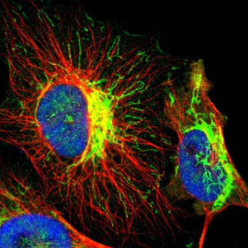
- Main image

- Experimental details
- Immunofluorescent staining of human cell line U-251 MG shows localization to mitochondria.
- Sample type
- Human