Explore
 Validate
 Learn
Learn Western blot
Antibody data
- Antibody Data
- Antigen structure
- References [3]
- Comments [0]
- Validations
- Western blot [2]
Submit
Validation data
Reference
Comment
Report error
- Product number
- PAB9930 - Provider product page

- Provider
- Abnova Corporation
- Proper citation
- Abnova Corporation Cat#PAB9930, RRID:AB_1676555
- Product name
- JUB polyclonal antibody
- Antibody type
- Polyclonal
- Description
- Rabbit polyclonal antibody raised against synthetic peptide of JUB.
- Storage
- Store at 4°C. For long term storage store at -20°C.Aliquot to avoid repeated freezing and thawing.
Submitted references Aurora-A and an interacting activator, the LIM protein Ajuba, are required for mitotic commitment in human cells.
The LIM protein Ajuba is recruited to cadherin-dependent cell junctions through an association with alpha-catenin.
Ajuba, a novel LIM protein, interacts with Grb2, augments mitogen-activated protein kinase activity in fibroblasts, and promotes meiotic maturation of Xenopus oocytes in a Grb2- and Ras-dependent manner.
Hirota T, Kunitoku N, Sasayama T, Marumoto T, Zhang D, Nitta M, Hatakeyama K, Saya H
Cell 2003 Sep 5;114(5):585-98
Cell 2003 Sep 5;114(5):585-98
The LIM protein Ajuba is recruited to cadherin-dependent cell junctions through an association with alpha-catenin.
Marie H, Pratt SJ, Betson M, Epple H, Kittler JT, Meek L, Moss SJ, Troyanovsky S, Attwell D, Longmore GD, Braga VM
The Journal of biological chemistry 2003 Jan 10;278(2):1220-8
The Journal of biological chemistry 2003 Jan 10;278(2):1220-8
Ajuba, a novel LIM protein, interacts with Grb2, augments mitogen-activated protein kinase activity in fibroblasts, and promotes meiotic maturation of Xenopus oocytes in a Grb2- and Ras-dependent manner.
Goyal RK, Lin P, Kanungo J, Payne AS, Muslin AJ, Longmore GD
Molecular and cellular biology 1999 Jun;19(6):4379-89
Molecular and cellular biology 1999 Jun;19(6):4379-89
No comments: Submit comment
Supportive validation
- Submitted by
- Abnova Corporation (provider)
- Main image
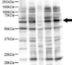
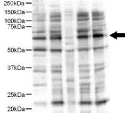
- Experimental details
- Western blot using JUB polyclonal antibody (Cat # PAB9930) shows detection of a 57 KDa band consistent with the expected MW for JUB (arrowhead).Lanes correspond to 1) HeLa nuclear extract, and 2) HeLa, 3) A-431, 4) Jurkat and 5) 293 whole cell lysates.Immunoprecipitation of JUB followed by western blotting may result in cleaner background staining.Approximately 5 ug ofeach preparation was run on a SDS-PAGE and transferred onto nitrocellulose followed by reaction with a 1 : 500 dilution of JUB polyclonal antibody.Detection occurred using a 1 : 5,000 dilution of HRP-labeled Donkey anti-Rabbit IgG for 1 hour at room temperature.Achemiluminescence system was used for signal detection using a 60-sec exposure time.Personal Communication. E. Pugacheva, Fox Chase Cancer Center, Philadelphia, PA.
- Submitted by
- Abnova Corporation (provider)
- Main image

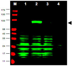
- Experimental details
- Western blot using JUB polyclonal antibody (Cat # PAB9930) shows detection of JUB-RFP fusion protein in cell lysates (arrow-head).Lanes correspond to 1) vector only transfection, 2) human JUB-RFP, 3) mouse JUB-RFP, and 4) mock transfection.Approximately 50 ug of each lysate was loaded per lane for SDS-PAGE followed by transfer onto nitrocellulose and reaction with a 1 : 1,700 dilution of JUB polyclonal antibody.Detection occurred using a 1 : 10,000 dilution of IRDye™800 conjugated Gt-a-Rabbit IgG [H&L] for 45 min at room temperature (800 nmchannel, green).Molecular weight estimation wasmade by comparison to prestained MW markers (indicated at left, 700 nm channel, red).IRDye™800 fluorescence image was captured using the Odyssey® Infrared Imaging System developed byLI-COR.IRDye is a trademark of LI-COR, Inc.