Explore
 Validate
 Learn
Learn Western blot
 ELISA
ELISAAntibody data
- Antibody Data
- Antigen structure
- References [0]
- Comments [0]
- Validations
- Western blot [2]
Submit
Validation data
Reference
Comment
Report error
- Product number
- AP09271PU-N - Provider product page

- Provider
- Acris Antibodies GmbH
- Proper citation
- Acris Antibodies GmbH Cat#AP09271PU-N, RRID:AB_2035885
- Product name
- anti Protein ajuba / JUB (224-239)
- Antibody type
- Polyclonal
- Antigen
- Synthetic peptide corresponding aa 224-239 of Human Ajuba
- Reactivity
- Human, Mouse, Rat, Canine
- Host
- Rabbit
- Isotype
- IgG
- Vial size
- 0.1 mg
- Concentration
- 1.67 mg/ml (by UV absorbance at 280 nm)
No comments: Submit comment
Supportive validation
- Submitted by
- Acris Antibodies GmbH (provider)
- Main image
- Experimental details
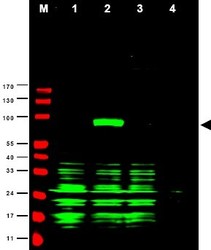
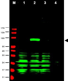
- Western blot using Affinity Purified anti-Ajuba antibody shows detection of Ajuba-RFP fusion protein in cell lysates (arrowhead). Lanes correspond to 1) vector only transfection, 2) human Ajuba-RFP, 3) mouse Ajuba-RFP, and 4) mock transfection. Approximately 50 µg of each lysate was loaded per lane for SDS-PAGE followed by transfer onto nitrocellulose and reaction with a 1:1,700 dilution of anti-Ajuba antibody. Detection occurred using a 1:10,000 dilution of IRDye(TM)800 conjugated Gt-a-Rabbit IgG [H&L] for 45 min at room temperature (800 nm channel, green). Molecular weight estimation was made by comparison to prestained MW markers (indicated at left, 700 nm channel, red). IRDye(TM)800 fluorescence image was captured using the Odyssey(R) Infrared Imaging System developed by LI-COR. IRDye is a trademark of LI-COR, Inc. Other detection systems will yield similar results.
- Submitted by
- Acris Antibodies GmbH (provider)
- Main image

- Experimental details
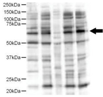
- Western blot using Affinity Purified anti-Ajuba antibody shows detection of a 57-kDa band consistent with the expected MW for Ajuba (arrowhead). Lanes correspond to 1) HeLa nuclear extract, and 2) HeLa, 3) A431, 4) Jurkat and 5) 293 whole cell lysates. Immunoprecipitation of Ajuba followed by western blotting may result in cleaner background staining. Approximately 5 µg of each preparation was run on a SDS-PAGE and transferred onto nitrocellulose followed by reaction with a 1:500 dilution of anti-Ajuba antibody. Detection occurred using a 1:5,000 dilution of HRP-labeled Donkey anti-Rabbit IgG for 1 hour at room temperature. A chemiluminescence system was used for signal detection (Roche) using a 60-sec exposure time.