Explore
 Validate
 Learn
Learn Western blot
 ELISA
ELISAAntibody data
- Antibody Data
- Antigen structure
- References [0]
- Comments [0]
- Validations
- Western blot [2]
Submit
Validation data
Reference
Comment
Report error
- Product number
- NB600-468 - Provider product page

- Provider
- Novus Biologicals
- Proper citation
- Novus Cat#NB600-468, RRID:AB_10003279
- Product name
- Rabbit Polyclonal Ajuba Antibody
- Antibody type
- Polyclonal
- Description
- Immunogen affinity purified. A BLAST analysis was used to suggest reactivity with this protein from human, rat, dog, mouse and chimpanzee based on 100% identity for the immunogen sequence. Cross reactivity with Ajuba protein homologues from other sources has not been determined.
- Reactivity
- Human, Mouse, Rat
- Host
- Rabbit
- Isotype
- IgG
- Vial size
- 0.1 mg
- Concentration
- 1.67 mg/ml
- Storage
- Store at -20C. Avoid freeze-thaw cycles.
No comments: Submit comment
Supportive validation
- Submitted by
- Novus Biologicals (provider)
- Main image
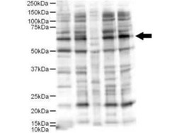
- Experimental details
- Western Blot: Ajuba Antibody [NB600-468] - Analysis of a 57-kDa band consistent with the expected MW for Ajuba (arrowhead). Lanes correspond to 1) HeLa nuclear extract, and 2) HeLa, 3) A431, 4) Jurkat and 5) 293 whole cell lysates. Immunoprecipitation of Ajuba followed by western blotting may result in cleaner background staining. Approximately 5 ug of each preparation was run on a SDS-PAGE and transferred onto nitrocellulose followed by reaction with a 1:500 dilution of anti-Ajuba antibody. Detection occurred using a 1:5,000 dilution of HRP-labeled Donkey anti-Rabbit IgG for 1 hour at room temperature. A chemiluminescence system was used for signal detection (Roche) using a 60-sec exposure time.

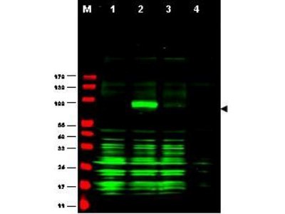
- Submitted by
- Novus Biologicals (provider)
- Main image
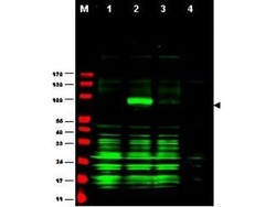
- Experimental details
- Western Blot: Ajuba Antibody [NB600-468] - Analysis of Ajuba-RFP fusion protein in cell lysates (arrow-head). Lanes correspond to 1) vector only trans-fection, 2) human Ajuba-RFP, 3) mouse Ajuba-RFP, and 4) mock transfection. Approximately 50 ug of each lysate was loaded per lane for SDS-PAGE followed by transfer onto nitrocellulose and reaction with a 1:1,700 dilution of anti-Ajuba antibody. Detection occurred using a 1:10,000 dilution of IRDye800 conjugated Gt-a-Rabbit IgG [H&L] for 45 min at room temperature (800 nm channel, green).