Explore
 Validate
 Learn
Learn Western blot
Antibody data
- Antibody Data
- Antigen structure
- References [0]
- Comments [0]
- Validations
- Western blot [2]
- Immunocytochemistry [1]
Submit
Validation data
Reference
Comment
Report error
- Product number
- 703805 - Provider product page

- Provider
- Invitrogen Antibodies
- Product name
- TMEM175 Recombinant Rabbit Monoclonal Antibody (5H18L65)
- Antibody type
- Monoclonal
- Antigen
- Synthetic peptide
- Reactivity
- Human
- Host
- Rabbit
- Isotype
- IgG
- Antibody clone number
- 5H18L65
- Vial size
- 100 µg
- Concentration
- 0.5 mg/mL
- Storage
- Store at 4°C short term. For long term storage, store at -20°C, avoiding freeze/thaw cycles.
No comments: Submit comment
Supportive validation
- Submitted by
- Invitrogen Antibodies (provider)
- Main image
- Experimental details
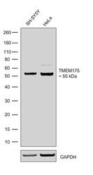
- Western blot was performed using Anti-TMEM175 Recombinant Rabbit Monoclonal Antibody (Product # 703805) and a 55 kDa band corresponding to TMEM175 was observed across the cell lines tested. Whole cell extracts (30 µg lysate) of SH-SY5Y (Lane 1) and HeLa (Lane 2) were electrophoresed using Novex® NuPAGE™ 4-12% Bis-Tris Protein Gel (Product # NP0321BOX). Resolved proteins were then transferred onto a nitrocellulose membrane (Product # IB23001) by iBlot® 2 Dry Blotting System (Product # IB21001). The blot was probed with the primary antibody (1:1000 dilution) and detected by chemiluminescence Goat Anti-Rabbit IgG Secondary Antibody, HRP conjugate (Product # A27036, 1:5000 dilution) using the iBright FL 1500 (Product # A44115). Chemiluminescent detection was performed using Novex® ECL Chemiluminescent Substrate Reagent Kit (Product # WP20005).
- Submitted by
- Invitrogen Antibodies (provider)
- Main image
- Experimental details
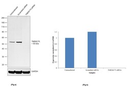
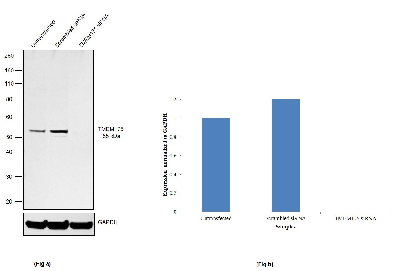
- Knockdown of TMEM175 was achieved by transfecting SH-SY5Y cells with TMEM175 specific siRNA (Silencer® select Product # s38819 & s38821). Western blot analysis (Fig a) was performed using whole cell extracts from TMEM175 knockdown cells (Lane 3), non-specific scrambled siRNA transfected cells (Lane 2) and untransfected cells (Lane 1). The blot was probed with Anti-TMEM175 Recombinant Rabbit Monoclonal Antibody (Product # 703805, 1:1000 dilution) and Goat anti-Rabbit IgG (H+L) Superclonal™ Secondary Antibody, HRP conjugate (Product # A27036, 1:5000 dilution). Densitometric analysis of this western blot is shown in the histogram (Fig b). Loss of signal upon siRNA mediated knockdown confirms that antibody is specific to TMEM175.
Supportive validation
- Submitted by
- Invitrogen Antibodies (provider)
- Main image
- Experimental details
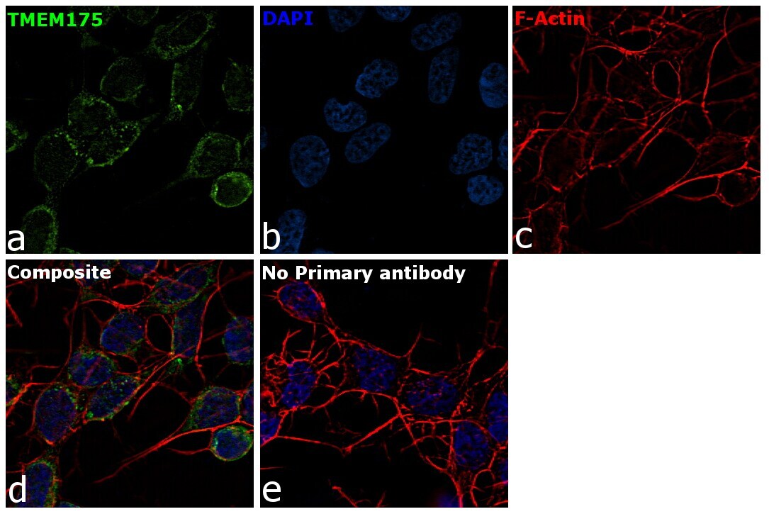
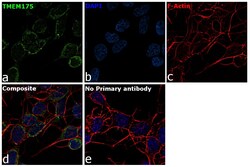
- For immunofluorescence analysis, SH-SY5Y cells were fixed and permeabilized for detection of endogenous TMEM175 using Anti-TMEM175 Recombinant Rabit Monoclonal Antibody (Product # 703805, 1:100 dilution) and labeled with Goat anti-Rabbit IgG (H+L) Highly Cross-Adsorbed Secondary Antibody, Alexa Fluor Plus 488 conjugate (Product # A32731, 1:2000). Panel a) shows representative cells that were stained for detection and localization of TMEM175 protein (green), Panel b) is stained for nuclei (blue) using ProLong™ Diamond Antifade Mountant with DAPI (Product # P36962). Panel c) represents cytoskeletal F-actin staining using Rhodamine Phalloidin (Product # R415, 1:300). Panel d) is a composite image of Panels a, b and c clearly demonstrating cytoplasmic and vesicular localization of TMEM175. Panel e) represents control cells with no primary antibody to assess background. The images were captured at 60X magnification.