Explore
 Validate
 Learn
LearnPA5-41325
antibody from Invitrogen Antibodies
Targeting: MCM8
C20orf154, dJ967N21.5, MGC119522, MGC119523, MGC12866, MGC4816, REC
 Western blot
Antibody data
- Antibody Data
- Antigen structure
- References [2]
- Comments [0]
- Validations
- Western blot [1]
- Immunohistochemistry [1]
- Other assay [2]
Submit
Validation data
Reference
Comment
Report error
- Product number
- PA5-41325 - Provider product page

- Provider
- Invitrogen Antibodies
- Product name
- MCM8 Polyclonal Antibody
- Antibody type
- Polyclonal
- Antigen
- Synthetic peptide
- Description
- Peptide sequence: IRLTEARARL ELREEATKED AEDIVEIMKY SMLGTYSDEF GNLDFERSQH Sequence homology: Cow: 100%; Dog: 93%; Guinea Pig: 100%; Horse: 100%; Human: 100%; Mouse: 100%; Rabbit: 100%; Rat: 100%; Zebrafish: 86%
- Reactivity
- Human
- Host
- Rabbit
- Isotype
- IgG
- Vial size
- 100 μL
- Concentration
- 0.5 mg/mL
- Storage
- -20°C, Avoid Freeze/Thaw Cycles
Submitted references Downregulation of MCM8 expression restrains the malignant progression of cholangiocarcinoma.
Identification of mini-chromosome maintenance 8 as a potential prognostic marker and its effects on proliferation and apoptosis in gastric cancer.
Hao J, Deng H, Yang Y, Chen L, Wu Q, Yao P, Li J, Li B, Jin X, Wang H, Duan H
Oncology reports 2021 Nov;46(5)
Oncology reports 2021 Nov;46(5)
Identification of mini-chromosome maintenance 8 as a potential prognostic marker and its effects on proliferation and apoptosis in gastric cancer.
Huang B, Lin M, Lu L, Chen W, Tan J, Zhao J, Cao Z, Zhu X, Lin J
Journal of cellular and molecular medicine 2020 Dec;24(24):14415-14425
Journal of cellular and molecular medicine 2020 Dec;24(24):14415-14425
No comments: Submit comment
Supportive validation
- Submitted by
- Invitrogen Antibodies (provider)
- Main image

- Experimental details
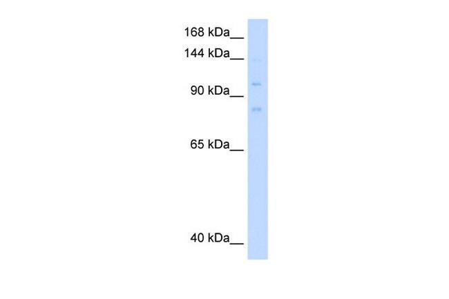
- Western blot analysis of human 721_B cell lysate using an anti-MCM8 polyclonal antibody (Product # PA5-41325).
Supportive validation
- Submitted by
- Invitrogen Antibodies (provider)
- Main image
- Experimental details
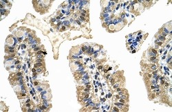
- Immunohistochemistry analysis of human intestine tissue using an anti-MCM8 polyclonal antibody (Product # PA5-41325).

Supportive validation
- Submitted by
- Invitrogen Antibodies (provider)
- Main image

- Experimental details
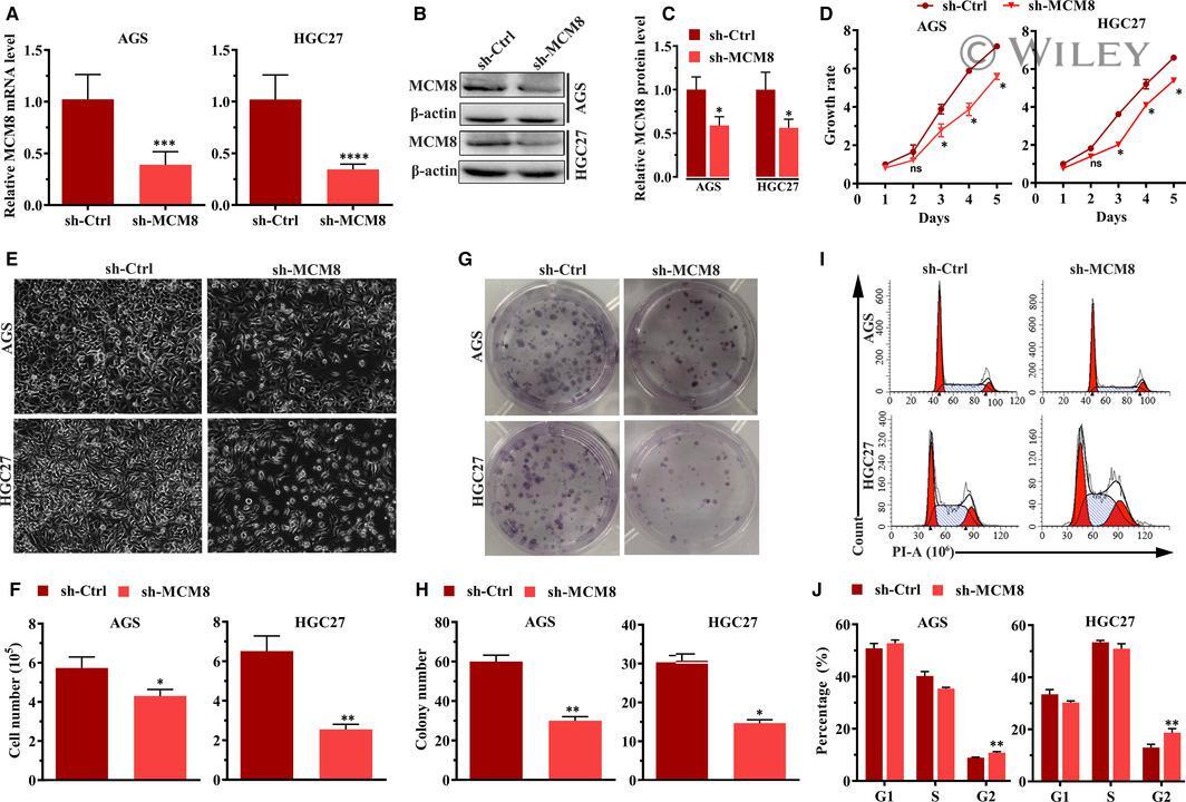
- 4 FIGURE MCM8 knockdown inhibits cell growth. A, MCM8 mRNA levels in AGS and HGC27 cells were detected by RT-qPCR. *** P < .001, **** P < .0001. B, Analysis of Western blotting showing that protein levels of MCM8 are reduced after MCM8 knockdown. C, Semi-quantification of Western blotting. The integrated band density was determined using the ImageLab Software, and beta-actin was used as the reference. * P < .05. D, MTT assay showing that MCM8 knockdown inhibits cell viability. ns: not significant. * P < .05. E, Morphological changes in AGS and HGC cells after infection with sh-MCM8 vs sh-Ctrl lentiviruses for 72 h. Cell morphology was observed under a phase-contrast microscope. Images were obtained at a magnification of 200x. F, MCM8 knockdown inhibits cell growth. * P < .05, ** P < .01. G, Representative images of colony formation assay. H, MCM8 knockdown reduces the number of colonies. * P < .05, ** P < .01. I, Representative images of cell cycle assay by flow cytometry. J, MCM8 knockdown induces arrest at the G2/M phase. ** P < .01. RT-qPCR, quantitative reverse transcription polymerase chain reaction
- Submitted by
- Invitrogen Antibodies (provider)
- Main image
- Experimental details
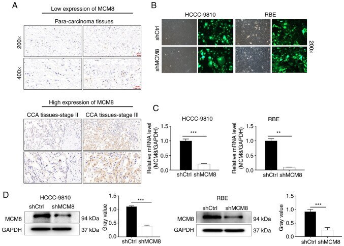
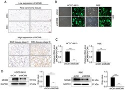
- Figure 1. Expression level of MCM8 in CCA. (A) The expression level of MCM8 in paracancerous tissues and tumor tissues of CCA patients was determined by immunohistochemical staining. (B) The efficiency of lentivirus transduction of HCCC-9810 and RBE cells was evaluated by the expression of green fluorescent protein. (C and D) The expression of MCM8 in HCCC-9810 and RBE cells after the stable transduction of lentivirus shMCM8 was assessed by (C) quantitative PCR and (D) western blot analysis. The data are presented as the mean +- SD (n=3). **P