 Explore
 Validate
 Learn
LearnMA5-34795
antibody from Invitrogen Antibodies
Targeting: CHAF1B
CAF-1, CAF1, CAF1A, CAF1P60, MPHOSPH7, MPP7
 Western blot
 Immunoprecipitation
Antibody data
- Antibody Data
- Antigen structure
- References [0]
- Comments [0]
- Validations
- Western blot [1]
- Immunocytochemistry [5]
- Immunohistochemistry [6]
- Flow cytometry [2]
Submit
Validation data
Reference
Comment
Report error
- Product number
- MA5-34795 - Provider product page

- Provider
- Invitrogen Antibodies
- Product name
- CAF1 p60 Recombinant Rabbit Monoclonal Antibody (JG92-30)
- Antibody type
- Monoclonal
- Antigen
- Recombinant full-length protein
- Description
- Positive Control: SiHa, K562, A431, 293T, human tonsil tissue, human appendix tissue, human colon tissue.
- Reactivity
- Human
- Host
- Rabbit
- Isotype
- IgG
- Antibody clone number
- JG92-30
- Vial size
- 100 μL
- Concentration
- 1 mg/mL
- Storage
- -20°C, Avoid Freeze/Thaw Cycles, store in dark
No comments: Submit comment
Supportive validation
- Submitted by
- Invitrogen Antibodies (provider)
- Main image

- Experimental details
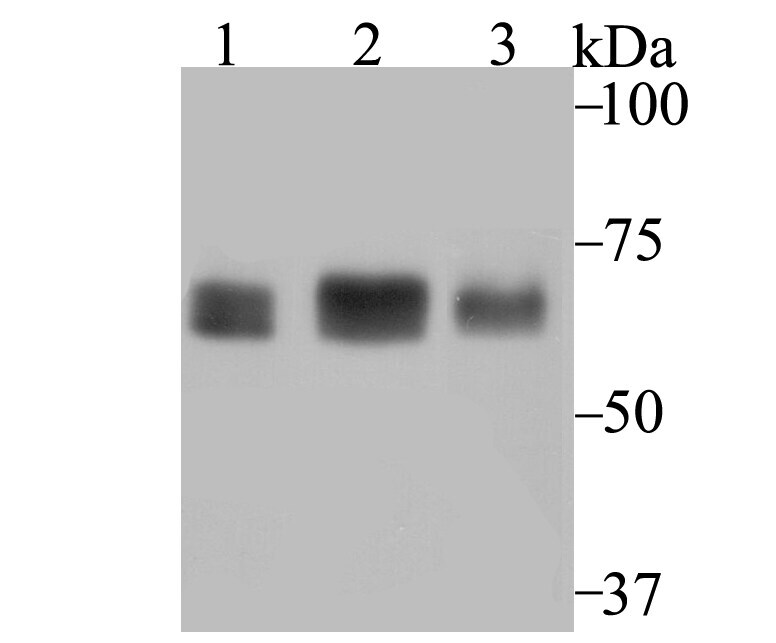
- Western blot analysis of p60 CAF1 in Positive control: Lane 1: SiHa, Lane 2: K562, Lane 3: A431. Samples were incubated with p60 CAF1 monoclonal antibody (Product # MA5-34795), at a dilution of 1:1000.
Supportive validation
- Submitted by
- Invitrogen Antibodies (provider)
- Main image
- Experimental details
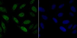
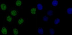
- Immunofluorescent analysis of p60 CAF1 in SiHa cells (green). Samples were fixed in paraformaldehyde and permeabilised with 0.25% Triton X100/PBS, incubated with p60 CAF1 monoclonal antibody (Product # MA5-34795), followed by DAPI (blue).
- Submitted by
- Invitrogen Antibodies (provider)
- Main image

- Experimental details
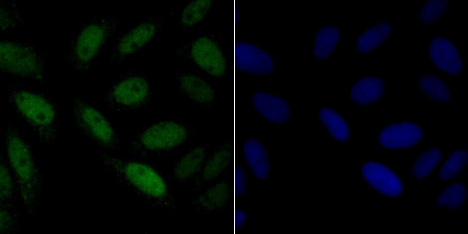
- Immunofluorescent analysis of p60 CAF1 in A431 cells (green). Samples were fixed in paraformaldehyde and permeabilised with 0.25% Triton X100/PBS, incubated with p60 CAF1 monoclonal antibody (Product # MA5-34795), followed by DAPI (blue).

- Submitted by
- Invitrogen Antibodies (provider)
- Main image
- Experimental details
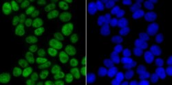
- Immunofluorescent analysis of p60 CAF1 in 293T cells (green). Samples were fixed in paraformaldehyde and permeabilised with 0.25% Triton X100/PBS, incubated with p60 CAF1 monoclonal antibody (Product # MA5-34795), followed by DAPI (blue).

- Submitted by
- Invitrogen Antibodies (provider)
- Main image

- Experimental details
- Immunofluorescent analysis of p60 CAF1 in 293T cells (green). Samples were fixed in paraformaldehyde and permeabilised with 0.25% Triton X100/PBS, incubated with p60 CAF1 monoclonal antibody (Product # MA5-34795), followed by DAPI (blue).

- Submitted by
- Invitrogen Antibodies (provider)
- Main image
- Experimental details
- Immunofluorescent analysis of p60 CAF1 in A431 cells (green). Samples were fixed in paraformaldehyde and permeabilised with 0.25% Triton X100/PBS, incubated with p60 CAF1 monoclonal antibody (Product # MA5-34795), followed by DAPI (blue).

Supportive validation
- Submitted by
- Invitrogen Antibodies (provider)
- Main image

- Experimental details
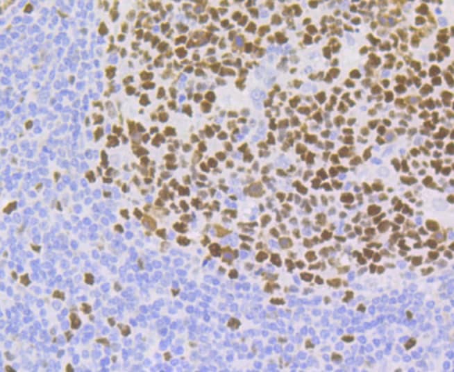
- Immunohistochemistry analysis of p60 CAF1 in paraffin-embedded human tonsil tissue. Samples were incubated with p60 CAF1 monoclonal antibody (Product # MA5-34795), and followed by hematoxylin.

- Submitted by
- Invitrogen Antibodies (provider)
- Main image
- Experimental details
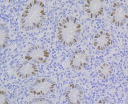
- Immunohistochemistry analysis of p60 CAF1 in paraffin-embedded human appendix tissue. Samples were incubated with p60 CAF1 monoclonal antibody (Product # MA5-34795), and followed by hematoxylin.
- Submitted by
- Invitrogen Antibodies (provider)
- Main image
- Experimental details
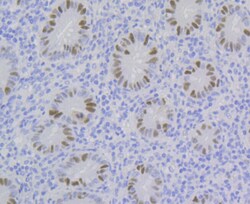
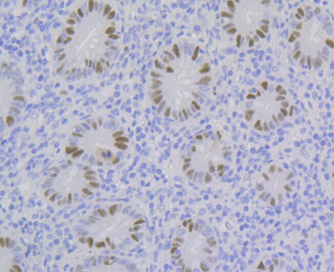
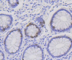
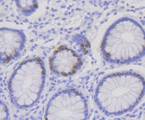
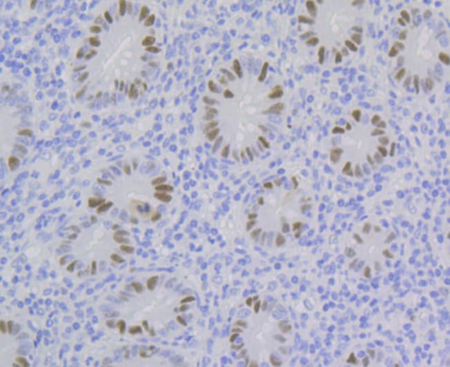
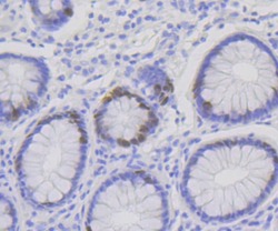
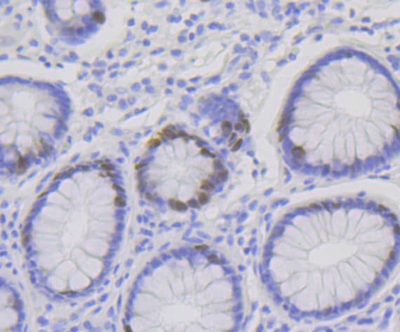
- Immunohistochemistry analysis of p60 CAF1 in paraffin-embedded human colon tissue. Samples were incubated with p60 CAF1 monoclonal antibody (Product # MA5-34795), and followed by hematoxylin.
- Submitted by
- Invitrogen Antibodies (provider)
- Main image

- Experimental details
- Immunohistochemistry analysis of p60 CAF1 in paraffin-embedded human tonsil tissue. Samples were incubated with p60 CAF1 monoclonal antibody (Product # MA5-34795), and followed by hematoxylin.
- Submitted by
- Invitrogen Antibodies (provider)
- Main image
- Experimental details
- Immunohistochemistry analysis of p60 CAF1 in paraffin-embedded human appendix tissue. Samples were incubated with p60 CAF1 monoclonal antibody (Product # MA5-34795), and followed by hematoxylin.
- Submitted by
- Invitrogen Antibodies (provider)
- Main image
- Experimental details
- Immunohistochemistry analysis of p60 CAF1 in paraffin-embedded human colon tissue. Samples were incubated with p60 CAF1 monoclonal antibody (Product # MA5-34795), and followed by hematoxylin.
Supportive validation
- Submitted by
- Invitrogen Antibodies (provider)
- Main image

- Experimental details
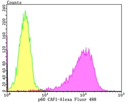
- Flow cytometry of p60 CAF1 in K562 cells (purple) compared with an unlabelled control (cells without incubation with primary antibody; yellow). Samples were incubated with p60 CAF1 monoclonal antibody (Product # MA5-34795) at a dilution of 1:100, followed by Alexa Fluor 488-conjugated goat anti-rabbit IgG.

- Submitted by
- Invitrogen Antibodies (provider)
- Main image
- Experimental details
- Flow cytometry of p60 CAF1 in K562 cells (purple) compared with an unlabelled control (cells without incubation with primary antibody; yellow). Samples were incubated with p60 CAF1 monoclonal antibody (Product # MA5-34795) at a dilution of 1:100, followed by Alexa Fluor 488-conjugated goat anti-rabbit IgG.
