Explore
 Validate
 Learn
Learn Western blot
Antibody data
- Antibody Data
- Antigen structure
- References [0]
- Comments [0]
- Validations
- Western blot [4]
- Immunocytochemistry [1]
Submit
Validation data
Reference
Comment
Report error
- Product number
- PA5-47914 - Provider product page

- Provider
- Invitrogen Antibodies
- Product name
- EMP Polyclonal Antibody
- Antibody type
- Polyclonal
- Antigen
- Recombinant full-length protein
- Description
- Reconstitute in sterile PBS to a final concentration of 0.2 mg/mL.
- Reactivity
- Human, Mouse, Rat
- Host
- Sheep
- Isotype
- IgG
- Vial size
- 100 µg
- Concentration
- 0.2 mg/mL
- Storage
- -20° C, Avoid Freeze/Thaw Cycles
No comments: Submit comment
Supportive validation
- Submitted by
- Invitrogen Antibodies (provider)
- Main image
- Experimental details
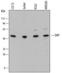
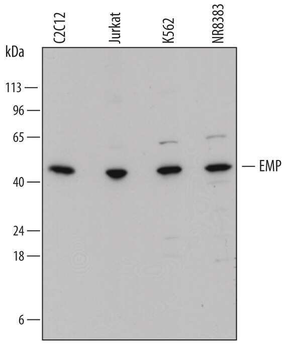
- Western blot analysis from lysates of C2C12 mouse myoblast cell line, Jurkat human acute T cell leukemia cell line, K562 human chronic myelogenous leukemia cell line, and NR8383 rat alveolar macrophage cell line. PVDF membrane was probed with 1 µg/mL of Sheep Anti-human EMP/MAEA Antigen Affinity-purified Polyclonal Antibody (Product # PA5-47914) followed by HRP-conjugated Anti-Sheep IgG Secondary Antibody. A specific band was detected for EMP/MAEA at approximately 45 kDa (as indicated). This experiment was conducted under reducing conditions.
- Submitted by
- Invitrogen Antibodies (provider)
- Main image
- Experimental details
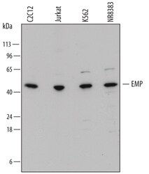
- Western blot analysis of EMP in C2C12 mouse myoblast cell line, Jurkat human acute T cell leukemia cell line, K562 human chronic myelogenous leukemia cell line, and NR8383 rat alveolar macrophage cell line. Samples were incubated in EMP polyclonal antibody (Product # PA5-47914) using a dilution of 1 µg/mL followed by a HRP-conjugated Anti-Sheep IgG secondary antibody. A specific band was detected for EMP/MAEA at approximately 45 kDa (as indicated). This experiment was conducted under reducing conditions.
- Submitted by
- Invitrogen Antibodies (provider)
- Main image

- Experimental details
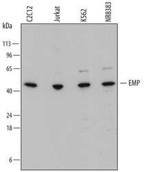
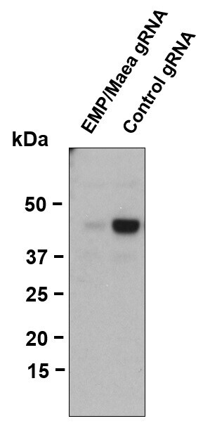
- Western blot analysis of EMP/MAEA was performed by loading whole cell lysate in 1X LDS sample buffer with 2-ME from 5 x 10⁵ Cas9-expressing mouse transformed pre-B cells. Samples were loaded onto a 4-12 % Bis-Tris polyacrylamide gel (Product # NP0336BOX). Proteins were transferred to nitrocellulose membrane by wet/tank transfer. Membrane was blocked in 5% milk/TBST. Target was detected at approximately 40 kDa using a polyclonal anti-EMP/Maea antibody (Product # PA5-47914) at a dilution of 0.5mg/ml in 5% milk/TBST at 4C overnight, followed by a secondary antibody HRP-anti-rabbit at a dilution of 1:5000 at room temperature for 1 hour. Chemiluminescent detection was performed using Pierce™ ECL Western Blotting Substrate (Product # PI-32209). Data courtesy of Thermo Scientific KOL Program.
- Submitted by
- Invitrogen Antibodies (provider)
- Main image
- Experimental details
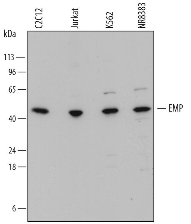
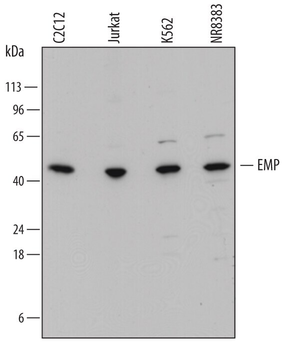
- Western blot analysis of EMP in C2C12 mouse myoblast cell line, Jurkat human acute T cell leukemia cell line, K562 human chronic myelogenous leukemia cell line, and NR8383 rat alveolar macrophage cell line. Samples were incubated in EMP polyclonal antibody (Product # PA5-47914) using a dilution of 1 µg/mL followed by a HRP-conjugated Anti-Sheep IgG secondary antibody. A specific band was detected for EMP/MAEA at approximately 45 kDa (as indicated). This experiment was conducted under reducing conditions.
Supportive validation
- Submitted by
- Invitrogen Antibodies (provider)
- Main image

- Experimental details
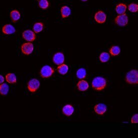
- Immunocytochemistry analysis of EMP in immersion fixed Jurkat human acute T cell leukemia cell line. Samples were incubated in EMP polyclonal antibody (Product # PA5-47914) using a dilution of 10 µg/mL for 3 hours at room temperature followed by NorthernLights™ 557-conjugated Anti-Sheep IgG Secondary Antibody (red) and counterstained with DAPI (blue). Specific staining was localized to cytoplasm.