Explore
 Validate
 Learn
Learn710862
antibody from Invitrogen Antibodies
Targeting: FERMT1
C20orf42, FLJ20116, KIND1, UNC112A, URP1
 Western blot
Antibody data
- Antibody Data
- Antigen structure
- References [0]
- Comments [0]
- Validations
- Western blot [3]
- Immunocytochemistry [1]
Submit
Validation data
Reference
Comment
Report error
- Product number
- 710862 - Provider product page

- Provider
- Invitrogen Antibodies
- Product name
- Kindlin Recombinant Polyclonal Antibody
- Antibody type
- Polyclonal
- Antigen
- Other
- Description
- This antibody is predicted to react with Monkey, Cat, Dog and Horse.
- Concentration
- 0.5 mg/mL
No comments: Submit comment
Supportive validation
- Submitted by
- Invitrogen Antibodies (provider)
- Main image
- Experimental details
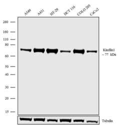
- Western blot analysis was performed on whole cell extracts (30 µg lysate) of A549 (Lane1), A431 (Lane 2), HT-29 (Lane 3), HCT116 (Lane 4), COLO-205 (Lane 5) and Caco-2 (Lane 6). The blots were probed with Anti-Kindlin1 Recombinant Rabbit Polyclonal Antibody (Product # 710862, 1-2 µg/mL) and detected by chemiluminescence using Goat anti-Rabbit IgG (H+L) Superclonal™ Secondary Antibody, HRP conjugate (Product # A27036, 0.4 µg/mL, 1:2500 dilution). A 77 kDa band corresponding to Kindlin1 was observed across cell lines tested. Known quantity of protein samples were electrophoresed using Novex® NuPAGE® 4-12% Bis-Tris gel (Product # NP0321BOX), XCell SureLock™ Electrophoresis System (Product # EI0002) and Novex® Sharp Pre-Stained Protein Standard (Product # LC5800). Resolved proteins were then transferred onto a nitrocellulose membrane with iBlot® Dry Blotting System (Product # IB21001). The membrane was probed with the relevant primary and secondary Antibody following blocking with 5% skimmed milk. Chemiluminescent detection was performed using Pierce™ ECL Western blotting Substrate (Product # 32106).

- Submitted by
- Invitrogen Antibodies (provider)
- Main image

- Experimental details
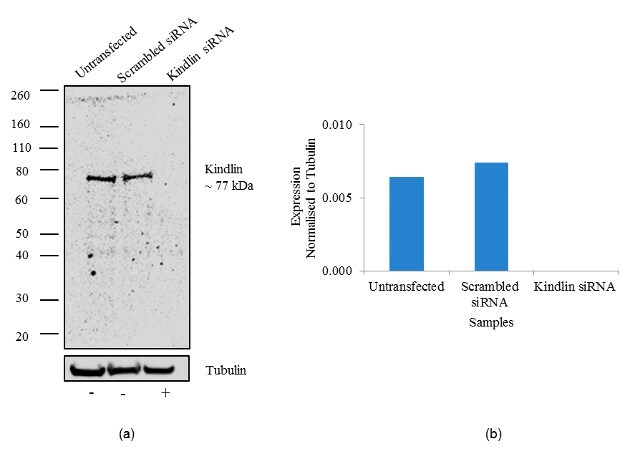
- Knockdown of was achieved by transfecting A549 cells with Kindlin 1 specific siRNA (Silencer® select Product # S31076 and s31074). Western blot analysis was performed using whole cell lysates from the Kindlin1 knock down cells (lane 3), non-specific scrambled siRNA transfected cells (lane 2) and untransfected cells (lane 1). The blots were probed with Anti-Kindlin1 Recombinant Rabbit Polyclonal Antibody (Product # 710862, 1-2 µg/mL) and detected by chemiluminescence using Goat anti-Rabbit IgG (H+L) Superclonal™ Secondary Antibody, HRP conjugate (Product # A27036, 0.4 µg/mL, 1:2500 dilution). Loss of signal upon siRNA mediated knock down confirms that antibody is specific to Kindlin1.
- Submitted by
- Invitrogen Antibodies (provider)
- Main image

- Experimental details
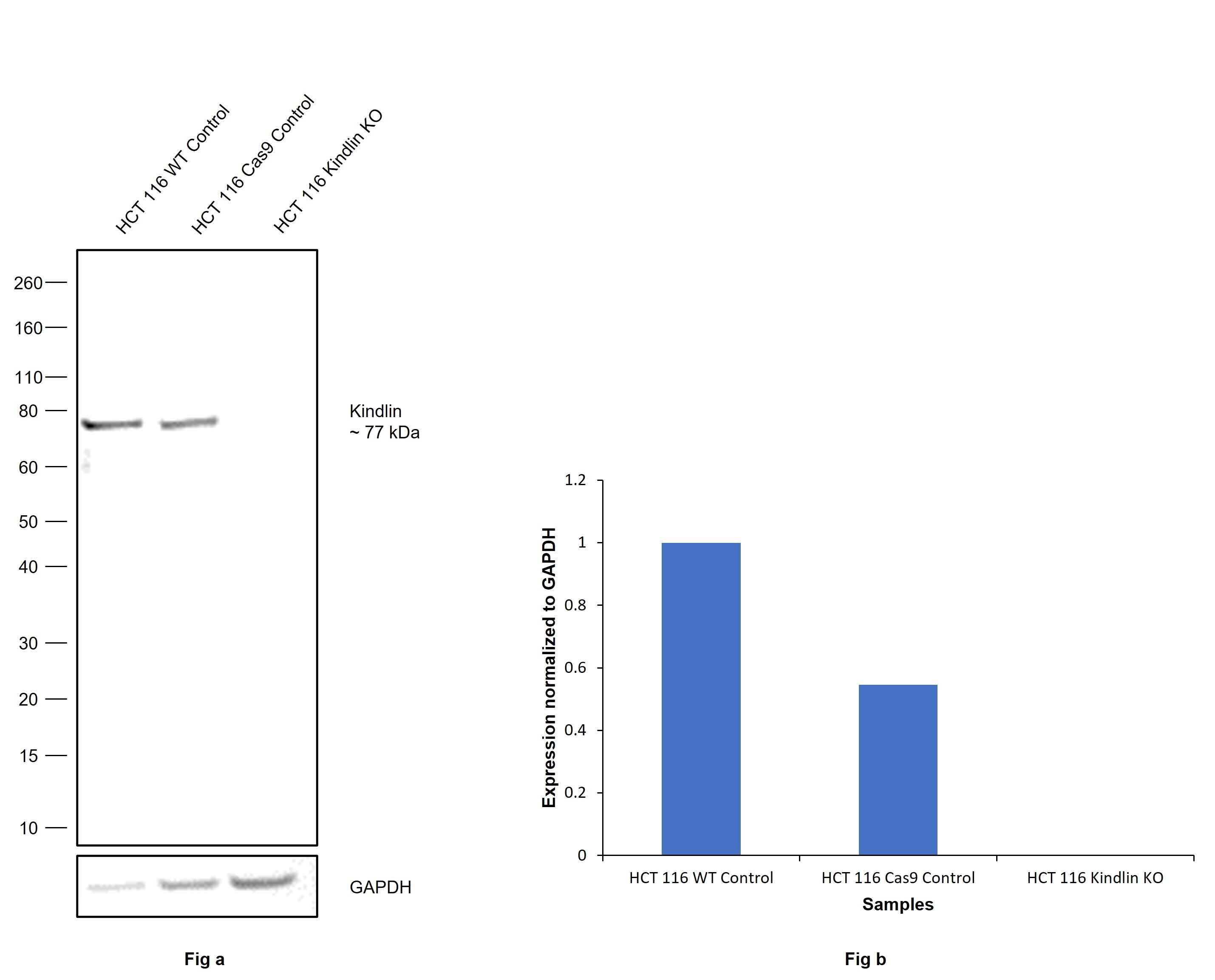
- Knockout of Kindlin was achieved by CRISPR-Cas9 genome editing using LentiArray™ Lentiviral sgRNA (Product # A32042, Assay ID CRIPSR637731_LV) and LentiArray Cas9 Lentivirus (Product # A32064). Western blot analysis of Kindlin was performed by loading 30 µg of HCT 116 wild type (Lane 1), HCT 116 Cas9 (Lane 2) andHCT 116 Kindlin KO (Lane 3) whole cell extracts. The samples were electrophoresed using NuPAGE™ Novex™ 4-12% Bis-Tris Protein Gel (Product # NP0322BOX). Resolved proteins were then transferred onto a nitrocellulose membrane (Product # IB23001) by iBlot® 2 Dry Blotting System (Product # IB21001). The blot was probed with Anti-Kindlin Recombinant Polyclonal Antibody (Product # 710862, 1:1000 dilution) and Goat anti-Rabbit IgG (H+L) Superclonal™ Recombinant Secondary Antibody, HRP (Product # A27036, 1:2500 dilution) using the iBright FL 1000 (Product # A32752). Chemiluminescent detection was performed using SuperSignal™ West Dura Extended Duration Substrate (Product # 34076). Loss of signal upon CRISPR mediated knockout (KO) using the LentiArray™ CRISPR product line confirms that antibody is specific to Kindlin.
Supportive validation
- Submitted by
- Invitrogen Antibodies (provider)
- Main image
- Experimental details
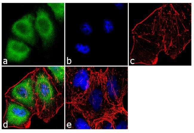
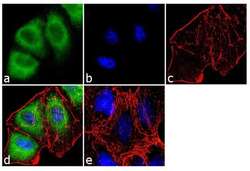
- Immunofluorescence was performed on fixed and permeabilized A-431 cells for detection of Kindlin1 using Kindlin1 Recombinant Rabbit Polyclonal Antibody (Product # 710862, 2 µg/mL) and labeled with Goat anti-Rabbit IgG (H+L) Superclonal™ Secondary Antibody, Alexa Fluor® 488 conjugate (Product # A27034, 1:2000). Panel a) shows representative cells that were stained for detection and localization of Kindlin1 protein (green), Panel b) is stained for nuclei (blue) using SlowFade® Gold Antifade Mountant with DAPI (Product # S36938). Panel c) represents cytoskeletal F-actin staining using Alexa Fluor® 555 Rhodamine Phalloidin (Product # R415, 1:300). Panel d) is a composite image of Panels a, b and c clearly demonstrating cytoplasmic localization of Kindlin1. Panel e) represents control cells with no primary antibody to assess background.