Explore
 Validate
 Learn
LearnPA3-16521
antibody from Invitrogen Antibodies
Targeting: HIF1A
bHLHe78, HIF-1alpha, HIF1, MOP1, PASD8
 Western blot
 ELISA Immunocytochemistry Immunoprecipitation Immunohistochemistry Gel shift Chromatin Immunoprecipitation Other assay
Antibody data
- Antibody Data
- Antigen structure
- References [4]
- Comments [0]
- Validations
- Western blot [5]
- Immunocytochemistry [2]
- Immunohistochemistry [3]
- Chromatin Immunoprecipitation [2]
- Other assay [2]
Submit
Validation data
Reference
Comment
Report error
- Product number
- PA3-16521 - Provider product page

- Provider
- Invitrogen Antibodies
- Product name
- HIF1A Polyclonal Antibody
- Antibody type
- Polyclonal
- Antigen
- Other
- Description
- For ChIP, nuclear extracts are recommended. Prior to immunostaining paraffin tissues, antigen retrieval with sodium citrate buffer (pH 6.0) is recommended. Suggested positive control: Cos-7 Hypoxia Induced nuclear extract Kit6.
- Reactivity
- Human, Mouse, Rat, Bovine, Canine, Guinea Pig, Hamster, Xenopus, Zebrafish
- Host
- Rabbit
- Isotype
- IgG
- Vial size
- 100 μL
- Concentration
- 1 mg/mL
- Storage
- Store at 4°C short term. For long term storage, store at -20°C, avoiding freeze/thaw cycles.
Submitted references α-Lipoic Acid Maintains Brain Glucose Metabolism via BDNF/TrkB/HIF-1α Signaling Pathway in P301S Mice.
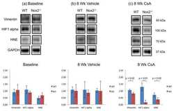
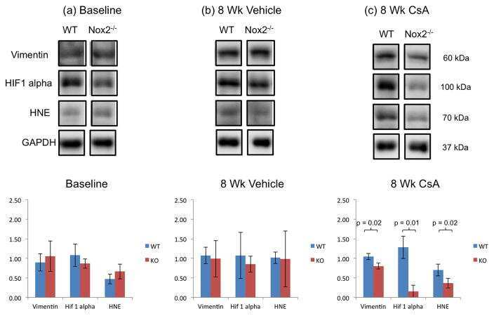
Nox2 and Cyclosporine-Induced Renal Hypoxia.
Dihydrotestosterone attenuates hypoxia inducible factor-1α and cyclooxygenase-2 in cerebral arteries during hypoxia or hypoxia with glucose deprivation.
Mouse snail is a target gene for HIF.
Zhang YH, Yan XZ, Xu SF, Pang ZQ, Li LB, Yang Y, Fan YG, Wang Z, Yu X, Guo C, Ao Q
Frontiers in aging neuroscience 2020;12:262
Frontiers in aging neuroscience 2020;12:262
Nox2 and Cyclosporine-Induced Renal Hypoxia.
Djamali A, Wilson NA, Sadowski EA, Zha W, Niles D, Hafez O, Dorn JR, Mehner TR, Grimm PC, Hoffmann FM, Zhong W, Fain SB, Reese SR
Transplantation 2016 Jun;100(6):1198-210
Transplantation 2016 Jun;100(6):1198-210
Dihydrotestosterone attenuates hypoxia inducible factor-1α and cyclooxygenase-2 in cerebral arteries during hypoxia or hypoxia with glucose deprivation.
Zuloaga KL, Gonzales RJ
American journal of physiology. Heart and circulatory physiology 2011 Nov;301(5):H1882-90
American journal of physiology. Heart and circulatory physiology 2011 Nov;301(5):H1882-90
Mouse snail is a target gene for HIF.
Luo D, Wang J, Li J, Post M
Molecular cancer research : MCR 2011 Feb;9(2):234-45
Molecular cancer research : MCR 2011 Feb;9(2):234-45
No comments: Submit comment
Supportive validation
- Submitted by
- Invitrogen Antibodies (provider)
- Main image
- Experimental details
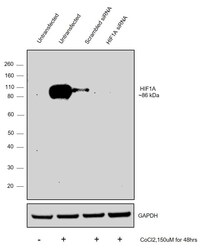
- Knockdown of HIF1A was achieved by transfecting MCF7 with HIF1A specific siRNAs (Silencer® select Product # S6541, S6539). Western blot analysis was performed using Nuclear enriched extracts from the HIF1A knockdown cells (treated with CoCl2, 150 µM for 48hrs) (lane 4), non-targeting scrambled siRNA transfected cells (treated with CoCl2, 150 µM for 48hrs) (lane 3), untransfected cells (treated with CoCl2, 150 µM for 48hrs) (lane 2) and untransfected cells (Lane 1). The blot was probed with HIF1A Polyclonal Antibody (Product # PA3-16521, 1:1000 dilution ) and Goat anti-Rabbit IgG (H+L) Superclonal™ Recombinant Secondary Antibody, HRP (Product # A27036, 1:20000 dilution). Decrease in signal upon siRNA mediated knock down confirms that antibody is specific to HIF1A.
- Submitted by
- Invitrogen Antibodies (provider)
- Main image
- Experimental details
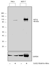
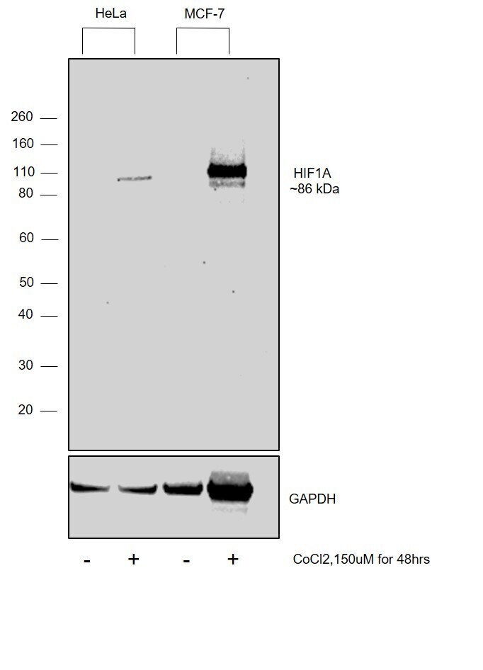
- Western blot was performed using Anti-HIF1A Polyclonal Antibody (Product # PA3-16521) and a 86 kDa band corresponding to HIF1A was observed across the panel upon induction with Cobalt Chloride. Nuclear enriched extracts (30 µg lysate) of HeLa (Lane 1), HeLa treated with CoCl2,150 µM for 48hrs (Lane 2), MCF7 (Lane 3), MCF7 treated with CoCl2,150 µM for 48hrs (Lane 4) were electrophoresed using NuPAGE™ 4-12% Bis-Tris Protein Gel (Product # NP0322BOX). Resolved proteins were then transferred onto a PVDF membrane (Product # IB23001) by iBlot® 2 Dry Blotting System (Product # IB21001). The blot was probed with the primary antibody (1:1000 dilution) and detected by chemiluminescence with Goat anti-Rabbit IgG (Heavy Chain) Superclonal™ Recombinant Secondary Antibody, HRP (Product # A27036,1:4000 dilution) using the iBright™ FL1500 Imaging System (Product # A44115). Chemiluminescentdetection was performed using Novex® ECL Chemiluminescent Substrate Reagent Kit (Product # WP20005).
- Submitted by
- Invitrogen Antibodies (provider)
- Main image

- Experimental details
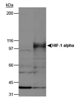
- Western blot analysis of HIF1A in normoxic and hypoxic nuclear rat cell lysates. Sample was incubated in HIF1A polyclonal antibody (Product # PA3-16521).
- Submitted by
- Invitrogen Antibodies (provider)
- Main image

- Experimental details
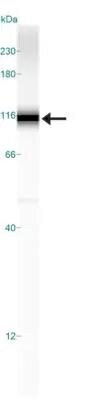
- Western blot analysis of HIF1A in 0.2 mg/mL Hypoxic HeLa lysate. Samples were incubated in HIF1A polyclonal antibody (Product # PA3-16521). This experiment was performed under reducing conditions using the 12-230 kDa separation system.
- Submitted by
- Invitrogen Antibodies (provider)
- Main image
- Experimental details
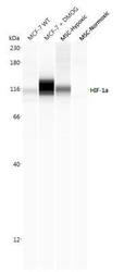
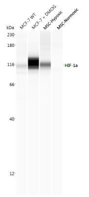
- Western blot analysis of HIF1A in 0.5 mg/mL lysates of MCF +/- DMOG, BioSpherix MSCs in hypoxic conditions, and BioSpherix MSCs in normoxic conditions. Samples were incubated in HIF1A polyclonal antibody (Product # PA3-16521) using a dilution of 1:100 followed by HRP-conjugated Anti-Rabbit. A band was detected for HIF-1 alpha at approximately 116 kDa (as indicated). This experiment was conducted under standard assay conditions, and using the 12-230 kDa separation module. Non-specific interaction with the 230 kDa Simple Western standard may be seen with this antibody.
Supportive validation
- Submitted by
- Invitrogen Antibodies (provider)
- Main image

- Experimental details
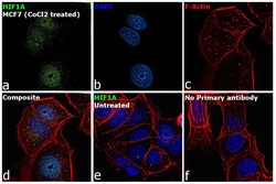
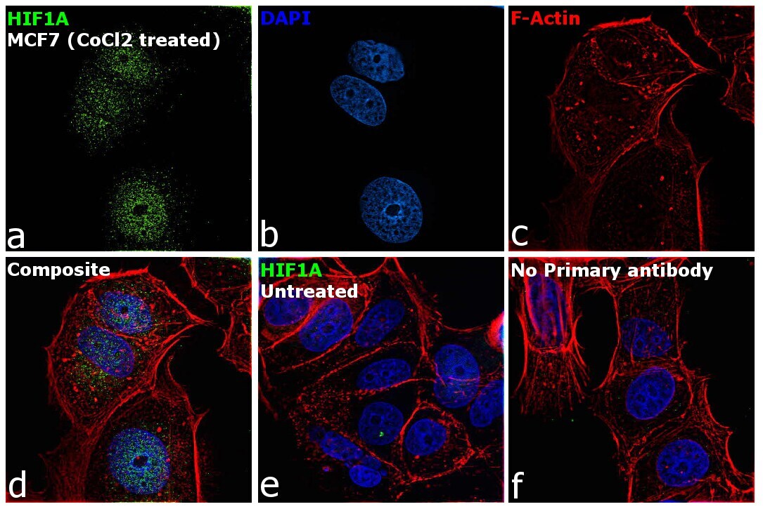
- Immunofluorescence analysis of HIF1A was performed using 70% confluent log phase MCF7 cells treated with Cobalt chloride (150 µM for 48hrs). The cells were fixed with 4% paraformaldehyde for 10 minutes, permeabilized with 0.1% Triton™ X-100 for 15 minutes and blocked with 2% BSA for 45 minutes at room temperature. The cells were labeled with HIF1A Polyclonal Antibody (Product # PA3-16521) at 1:200 dilution in 0.1% BSA, incubated at 4 degree celsius overnight and then labeled with Donkey anti-Rabbit IgG (H+L) Highly Cross-Adsorbed Secondary Antibody, Alexa Fluor Plus 488 (Product # A32790), (1:3000 dilution), for 45 minutes at room temperature (Panel a: Green). Nuclei (Panel b:Blue) were stained with ProLong™ Diamond Antifade Mountant with DAPI (Product # P36962). F-actin (Panel c: Red) was stained with Rhodamine Phalloidin (Product # R415, 1:300). Panel d represents the merged image showing nucleus localization in CoCl2 treated MCF7 cells. Panel e represents no staining observed for untreated MCF7 cells. Panel f represents control cells with no primary antibody to assess background. The images were captured at 60X magnification.

- Submitted by
- Invitrogen Antibodies (provider)
- Main image
- Experimental details
- Immunofluorescence analysis of HIF1A was performed using 70% confluent log phase MCF7 cells treated with Cobalt chloride (150 µM for 48hrs). The cells were fixed with 4% paraformaldehyde for 10 minutes, permeabilized with 0.1% Triton™ X-100 for 15 minutes and blocked with 2% BSA for 45 minutes at room temperature. The cells were labeled with HIF1A Polyclonal Antibody (Product # PA3-16521) at 1:200 dilution in 0.1% BSA, incubated at 4 degree celsius overnight and then labeled with Donkey anti-Rabbit IgG (H+L) Highly Cross-Adsorbed Secondary Antibody, Alexa Fluor Plus 488 (Product # A32790), (1:3000 dilution), for 45 minutes at room temperature (Panel a: Green). Nuclei (Panel b:Blue) were stained with ProLong™ Diamond Antifade Mountant with DAPI (Product # P36962). F-actin (Panel c: Red) was stained with Rhodamine Phalloidin (Product # R415, 1:300). Panel d represents the merged image showing nucleus localization in CoCl2 treated MCF7 cells. Panel e represents no staining observed for untreated MCF7 cells. Panel f represents control cells with no primary antibody to assess background. The images were captured at 60X magnification.
Supportive validation
- Submitted by
- Invitrogen Antibodies (provider)
- Main image
- Experimental details
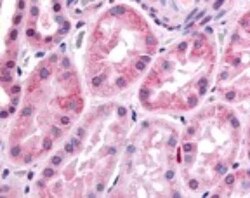
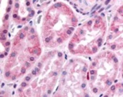
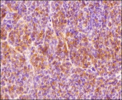
- Immunohistochemical analysis of HIF1A in human kidney, renal tubular epithelium in cortex. Samples were incubated in HIF1A polyclonal antibody (Product # PA3-16521).
- Submitted by
- Invitrogen Antibodies (provider)
- Main image
- Experimental details
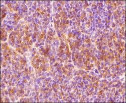
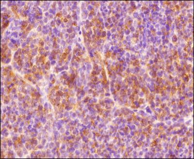
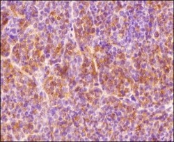
- Immunohistochemical analysis of HIF1A in formalin-fixed paraffin-embedded tissue section of human endometrium carcinoma AN3CA cell line based xenograft. Samples were incubated in HIF1A polyclonal antibody (Product # PA3-16521) using a dilution of 1:300 followed by a HRP-labeled secondary antibody and DAB reagent. The section was further counterstained using hematoxylin. The tested section depicted mainly a diffused cytoplasmic staining but there were some cells which showed nuclear signal also (representing hypoxic cells).
- Submitted by
- Invitrogen Antibodies (provider)
- Main image
- Experimental details
- Immunohistochemical analysis of HIF1A in formalin-fixed paraffin-embedded tissue section of human endometrium carcinoma AN3CA cell line based xenograft. Samples were incubated in HIF1A polyclonal antibody (Product # PA3-16521) using a dilution of 1:300 followed by a HRP-labeled secondary antibody and DAB reagent. The section was further counterstained using hematoxylin. The tested section depicted mainly a diffused cytoplasmic staining but there were some cells which showed nuclear signal also (representing hypoxic cells).
Supportive validation
- Submitted by
- Invitrogen Antibodies (provider)
- Main image
- Experimental details
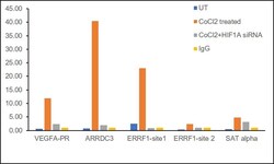
- Chromatin Immunoprecipitation (ChIP) assay of endogenous HIF1A was performed using HIF1A Polyclonal Antibody (Product # PA3-16521, 5 µg) on sheared chromatin from MCF-7 cells treated with Cobalt Chloride (150 µM for 48hrs) using the MAGnify ChIP System kit (Product # 49-2024). Chromatin from untreated MCF-7 cells were prepared the same way. Normal Rabbit IgG was used as a negative IP control. The purified DNA was analyzed by qPCR using primers binding to VEGFA-PR, ARRDC3, ERRF1-site 1 (active) and ERRF1-site 2 and SAT alpha (inactive). Data is presented as fold enrichment of the antibody signal versus the negative control IgG using the comparative CT method.

- Submitted by
- Invitrogen Antibodies (provider)
- Main image

- Experimental details
- Chromatin Immunoprecipitation (ChIP) assay of endogenous HIF1A was performed using HIF1A Polyclonal Antibody (Product # PA3-16521, 5 µg) on sheared chromatin from MCF-7 cells treated with Cobalt Chloride (150 µM for 48hrs) using the MAGnify ChIP System kit (Product # 49-2024). Chromatin from untreated MCF-7 cells were prepared the same way. Normal Rabbit IgG was used as a negative IP control. The purified DNA was analyzed by qPCR using primers binding to VEGFA-PR, ARRDC3, ERRF1-site 1 (active) and ERRF1-site 2 and SAT alpha (inactive). Data is presented as fold enrichment of the antibody signal versus the negative control IgG using the comparative CT method.

Supportive validation
- Submitted by
- Invitrogen Antibodies (provider)
- Main image
- Experimental details
- NULL
- Submitted by
- Invitrogen Antibodies (provider)
- Main image
- Experimental details
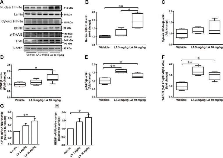
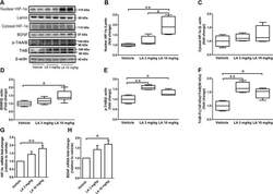
- Figure 3 LA mediated the upregulation of hypoxia-inducible factor-1alpha (HIF-1alpha) expression via brain-derived neurotrophic factor/tyrosine Kinase receptor B (BDNF/TrkB) pathway. (A) Western blot results showed the protein expression levels of nuclear HIF-1alpha, Lamin, and cytosol HIF-1alpha, BDNF, p-TrkA/B, TrkB. (B) Quantified results of nuclear HIF-1alpha levels. Lamin served as an internal loading control. (C-F) Relative protein levels of cytosol HIF-1alpha, BDNF, p-TrkB, TrkB-FL which were established after normalization to beta-actin. (G,H) mRNA levels of HIF-1alpha and BDNF. Values are represented as the means +- SEM ( n = 7). * p < 0.05, ** p < 0.01 compared with the vehicle group.