 Explore
 Validate
 Learn
Learn Western blot
 Immunocytochemistry
Antibody data
- Antibody Data
- Antigen structure
- References [0]
- Comments [0]
- Validations
- Western blot [1]
- Immunohistochemistry [3]
Submit
Validation data
Reference
Comment
Report error
- Product number
- LS-C100908 - Provider product page

- Provider
- LSBio
- Proper citation
- LifeSpan Cat#LS-C100908, RRID:AB_2282386
- Product name
- MSRA Antibody (aa34-63) LS-C100908
- Antibody type
- Polyclonal
- Description
- Ammonium sulfate precipitation
- Reactivity
- Human, Mouse
- Host
- Rabbit
- Storage
- Maintain refrigerated at 2°C to 8°C for up to 6 months. For long term storage store at -20°C.
No comments: Submit comment
Enhanced validation
- Submitted by
- LSBio (provider)
- Enhanced method
- Genetic validation
- Main image

- Experimental details
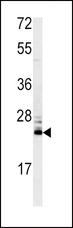
- Western blot of MSRA antibody in mouse kidney tissue lysates (35 ug/lane). MSRA (arrow) was detected using the purified antibody.
Enhanced validation
- Submitted by
- LSBio (provider)
- Enhanced method
- Genetic validation
- Main image
- Experimental details
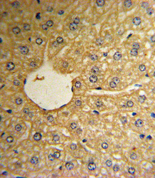
- Formalin-fixed and paraffin-embedded human hepatocarcinoma reacted with MSRA Antibody , which was peroxidase-conjugated to the secondary antibody, followed by DAB staining. This data demonstrates the use of this antibody for immunohistochemistry; clinical relevance has not been evaluated.

- Submitted by
- LSBio (provider)
- Enhanced method
- Genetic validation
- Main image

- Experimental details
- Formalin-fixed and paraffin-embedded human hepatocarcinoma reacted with MSRA Antibody , which was peroxidase-conjugated to the secondary antibody, followed by DAB staining. This data demonstrates the use of this antibody for immunohistochemistry; clinical relevance has not been evaluated.

- Submitted by
- LSBio (provider)
- Main image

- Experimental details
- Formalin-fixed and paraffin-embedded human hepatocarcinoma reacted with MSRA Antibody , which was peroxidase-conjugated to the secondary antibody, followed by DAB staining. This data demonstrates the use of this antibody for immunohistochemistry; clinical relevance has not been evaluated.
