Explore
 Validate
 Learn
Learn Western blot
Antibody data
- Antibody Data
- Antigen structure
- References [0]
- Comments [0]
- Validations
- Western blot [1]
- Immunohistochemistry [2]
Submit
Validation data
Reference
Comment
Report error
- Product number
- APR-013-200UL - Provider product page

- Provider
- Invitrogen Antibodies
- Product name
- P2X6 Receptor Polyclonal Antibody
- Antibody type
- Polyclonal
- Antigen
- Other
- Reactivity
- Human, Mouse, Rat
- Host
- Rabbit
- Isotype
- IgG
- Vial size
- 200 µL
- Concentration
- 0.7 mg/mL
- Storage
- -20° C, Avoid Freeze/Thaw Cycles
No comments: Submit comment
Supportive validation
- Submitted by
- Invitrogen Antibodies (provider)
- Main image

- Experimental details
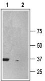
- Western blot analysisof rat brain membranes: - 1. Anti-P2X6 Receptor Antibody (#APR-013), (1:200). 2. Anti-P2X6 Receptor Antibody , preincubated with P2X6 Receptor Blocking Peptide (#BLP-PR013).
Supportive validation
- Submitted by
- Invitrogen Antibodies (provider)
- Main image
- Experimental details
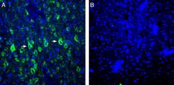
- Expression of P2X6 in rat parietal cortex. Immunohistochemical staining of perfusion-fixed frozen rat brain sections Anti-P2X6 Receptor Antibody (#APR-013), (1:200), followed by goat Anti-rabbit-AlexaFluor-488. A. P2X6 immunoreactivity (green) appears in cortical neurons (arrows). B. Pre-incubation of the Antibody with P2X6 Blocking Peptide (BLP-PR013), suppressed staining. Cell nuclei are stained with DAPI (blue).
- Submitted by
- Invitrogen Antibodies (provider)
- Main image
- Experimental details
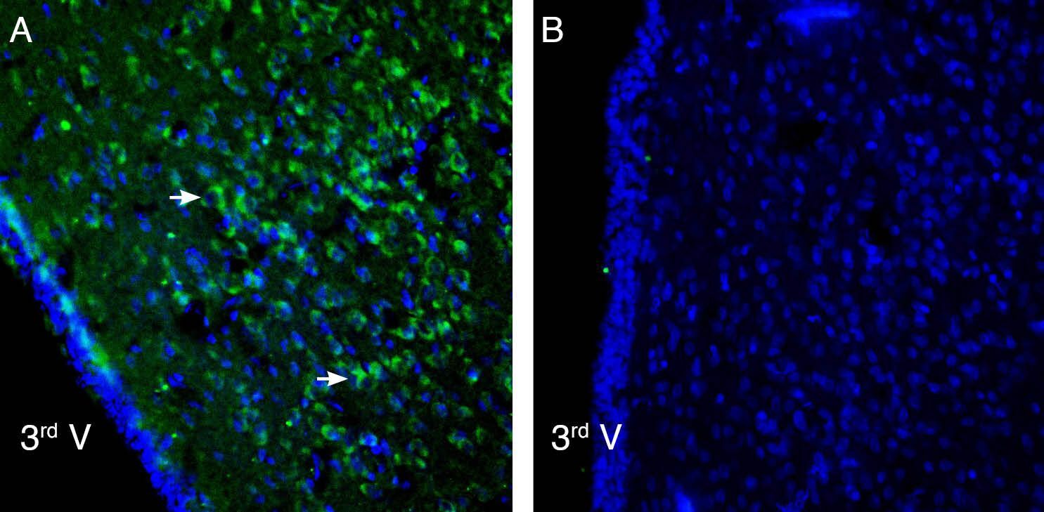
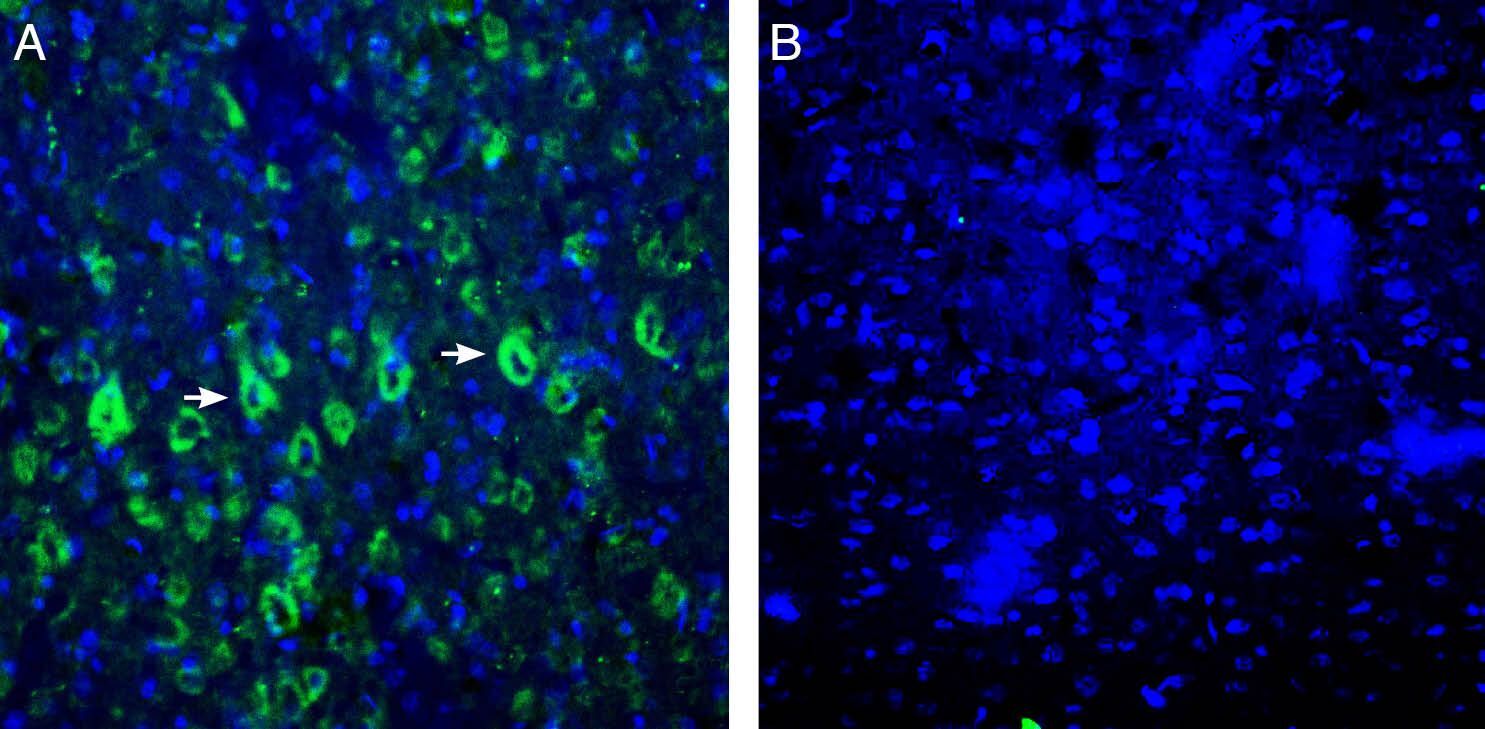
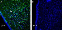
- Expression of P2X6 in rat hypothalamus. Immunohistochemical staining of perfusion-fixed frozen rat brain sections Anti-P2X6 Receptor Antibody (#APR-013), (1:200), followed by goat Anti-rabbit-AlexaFluor-488. A. P2X6 immunoreactivity (green) appears in neurons (arrows). B. Pre-incubation of the Antibody with P2X6 Blocking Peptide (BLP-PR013), suppressed staining. Cell nuclei are stained with DAPI (blue). 3rd V = Third ventricle.