 Explore
 Validate
 Learn
Learn Western blot
 Immunocytochemistry
Antibody data
- Antibody Data
- Antigen structure
- References [1]
- Comments [0]
- Validations
- Immunocytochemistry [1]
- Other assay [1]
Submit
Validation data
Reference
Comment
Report error
- Product number
- 710022 - Provider product page

- Provider
- Invitrogen Antibodies
- Product name
- BMP-2 Recombinant Superclonal™ Antibody (18HCLC)
- Antibody type
- Other
- Antigen
- Other
- Description
- This antibody is predicted to react with mouse based on sequence homology. Recombinant rabbit Superclonal™ antibodies are unique offerings from Thermo Fisher Scientific. They are comprised of a selection of multiple different recombinant monoclonal antibodies, providing the best of both worlds - the sensitivity of polyclonal antibodies with the specificity of monoclonal antibodies - all delivered with the consistency only found in a recombinant antibody. While functionally the same as a polyclonal antibody - recognizing multiple epitope sites on the target and producing higher detection sensitivity for low abundance targets - a recombinant rabbit Superclonal™ antibody has a known mixture of light and heavy chains. The exact population can be produced in every lot, circumventing the biological variability typically associated with polyclonal antibody production. Note: Formerly called “Recombinant polyclonal antibody”, this product is now rebranded as “Recombinant Superclonal™ antibody”. The physical product and the performance remain unchanged.
- Reactivity
- Human
- Host
- Rabbit
- Isotype
- IgG
- Antibody clone number
- 18HCLC
- Vial size
- 100 μg
- Concentration
- 0.5 mg/mL
- Storage
- Store at 4°C short term. For long term storage, store at -20°C, avoiding freeze/thaw cycles.
Submitted references The tyrosine kinase inhibitor nilotinib targets the discoidin domain receptor DDR2 in calcific aortic valve stenosis.
Carracedo M, Pawelzik SC, Artiach G, Pouwer MG, Plunde O, Saliba-Gustafsson P, Ehrenborg E, Eriksson P, Pieterman E, Stenke L, Princen HMG, Franco-Cereceda A, Bäck M
British journal of pharmacology 2022 Oct;179(19):4709-4721
British journal of pharmacology 2022 Oct;179(19):4709-4721
No comments: Submit comment
Supportive validation
- Submitted by
- Invitrogen Antibodies (provider)
- Main image
- Experimental details
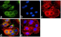
- Immunofluorescence analysis of BMP-2 was done on 70% confluent log phase MCF7 cells. The cells were fixed with 4% paraformaldehyde for 15 minutes, permeabilized with 0.25% Triton™ X-100 for 10 minutes, and blocked with 5% BSA for 1 hour at room temperature. The cells were labeled with BMP-2 (18HCLC), Recombinant Rabbit Superclonal™ Antibody (Product # 710022) at 1 µg/mL in 1% BSA and incubated for 3 hours at room temperature and then labeled with Goat anti-Rabbit IgG (Heavy Chain) Superclonal™ Secondary Antibody, Alexa Fluor® 488 conjugate (Product # A27034) at a dilution of 1:2000 for 45 minutes at room temperature (Panel a: green). Nuclei (Panel b: blue) were stained with SlowFade® Gold Antifade Mountant with DAPI (Product # S36938). F-actin (Panel c: red) was stained with Alexa Fluor® 555 Rhodamine Phalloidin (Product # R415, 1:300). Panel d is a merged image showing Cytoplasmic localization. Panel e is a no primary antibody control. The images were captured at 60X magnification.
Supportive validation
- Submitted by
- Invitrogen Antibodies (provider)
- Main image
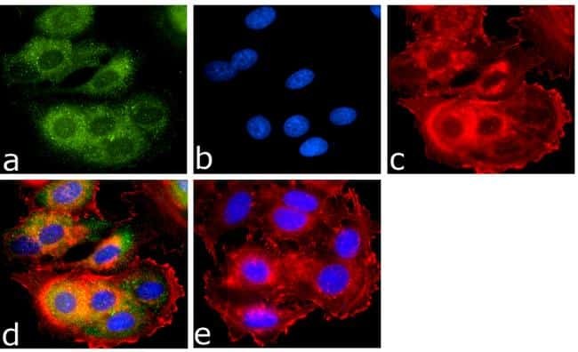
- Experimental details
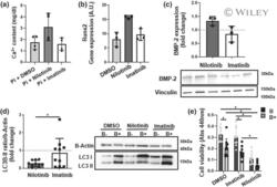
- 2 FIGURE Effects of nilotinib on human valvular interstitial cells. (a) Quantification of phosphate-induced calcification of human valvular interstitial cells (VICs) after 9 days. (b) mRNA expression of the osteogenic transcription factor RUNx-2 in VICs treated for 24 h. (c) Immunoblotting against BMP-2 in human VICs after 24 h treatment with control (DMSO), nilotinib (10 muM), or imatinib (10 muM). (d) Immunoblotting against LC3-I and LC3-II in human VICs treated for 2 h with or without bafilomycin (100 nM) followed by 24 h with control (DMSO), nilotinib (10 muM), or imatinib (10 muM). (e) VIC viability after inhibition of autophagy with bafilomycin (B+) (100 nM) or vehicle control (B-) (DMSO) for 2 h, followed by treatment with control (DMSO), nilotinib (10 muM) or imatinib (10 muM) for 24 h. Data are represented as mean +- S.D. Statistical significance of differences between groups were determined when at least n = 5 (d and e). * P < 0.05 Student's t test. Each dot represents one patient VIC donor
