Explore
 Validate
 Learn
Learn Western blot
 Immunohistochemistry
Antibody data
- Antibody Data
- Antigen structure
- References [1]
- Comments [0]
- Validations
- Immunohistochemistry [1]
- Other assay [3]
Submit
Validation data
Reference
Comment
Report error
- Product number
- PA5-42007 - Provider product page

- Provider
- Invitrogen Antibodies
- Product name
- PRPS2 Polyclonal Antibody
- Antibody type
- Polyclonal
- Antigen
- Synthetic peptide
- Description
- Peptide sequence: VSPDAGGAKR VTSIADRLNV EFALIHKERK KANEVDRMVL VGDVKDRVAI Sequence homology: Cow: 100%; Dog: 100%; Guinea Pig: 100%; Horse: 100%; Human: 100%; Mouse: 100%; Rabbit: 100%; Rat: 100%; Zebrafish: 100%
- Reactivity
- Human
- Host
- Rabbit
- Isotype
- IgG
- Vial size
- 100 μL
- Concentration
- 0.5 mg/mL
- Storage
- -20°C, Avoid Freeze/Thaw Cycles
Submitted references Phosphoribosyl-pyrophosphate synthetase 2 (PRPS2) depletion regulates spermatogenic cell apoptosis and is correlated with hypospermatogenesis.
Lei B, Xie LX, Zhang SB, Wan B, Zhong LR, Zhou XM, Mao XM, Shu FP
Asian journal of andrology 2020 Sep-Oct;22(5):493-499
Asian journal of andrology 2020 Sep-Oct;22(5):493-499
No comments: Submit comment
Supportive validation
- Submitted by
- Invitrogen Antibodies (provider)
- Main image
- Experimental details
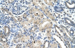
- Immunohistochemistry (paraffin-embedded) analysis of human kidney tissue using an anti-PRPS2 polyclonal antibody (Product # PA5-42007).

Supportive validation
- Submitted by
- Invitrogen Antibodies (provider)
- Main image
- Experimental details
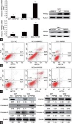
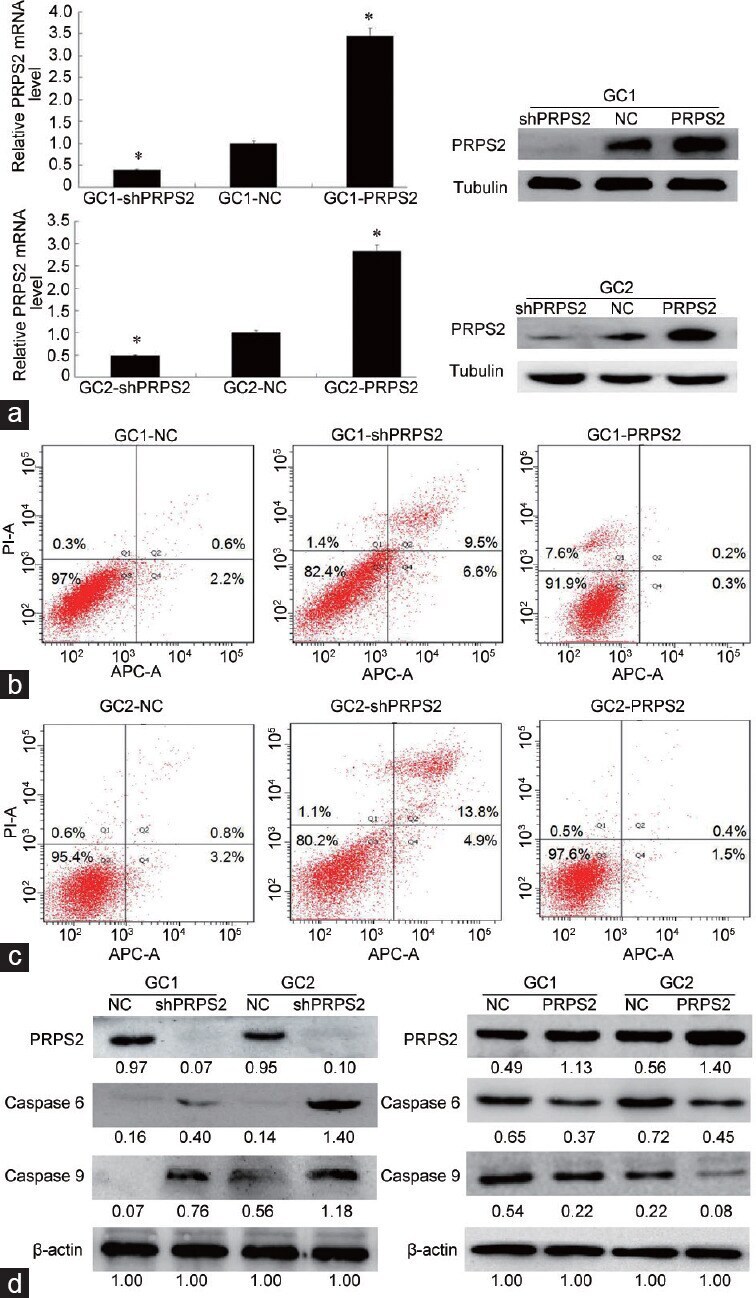
- Figure 1 PRPS2 depletion and apoptosis of spermatogenic cells. ( a ) qRT-PCR and Western blot analysis confirmed that PRPS2 was successfully downregulated by gene silencing and upregulated by PRPRS2 overexpression in both GC1 and GC2 cells. ( b ) Flow cytometry analysis shows that PRPS2 depletion promotes the apoptosis of GC1 cells. ( c ) Flow cytometry analysis shows that PRPS2 depletion promotes the apoptosis of GC2 cells (abscissa: cell count; ordinate: the fluorescence intensity). ( d ) Western blot analysis indicates that PRPS2 depletion activates the expression of apoptotic proteins (values beneath blots are relative to the control). *Significantly different compared with negative control, P < 0.05 by independent-samples t -test. shPRPS2: small-hairpin RNA gene silencer; NC: negative control; PRPS2: overexpression; PRPS2: phosphoribosyl-pyrophosphate synthetase 2; qRT-PCR: quantitative real-time polymerase chain reaction; APC-A: Allophycocyanin-A.
- Submitted by
- Invitrogen Antibodies (provider)
- Main image
- Experimental details
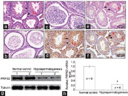
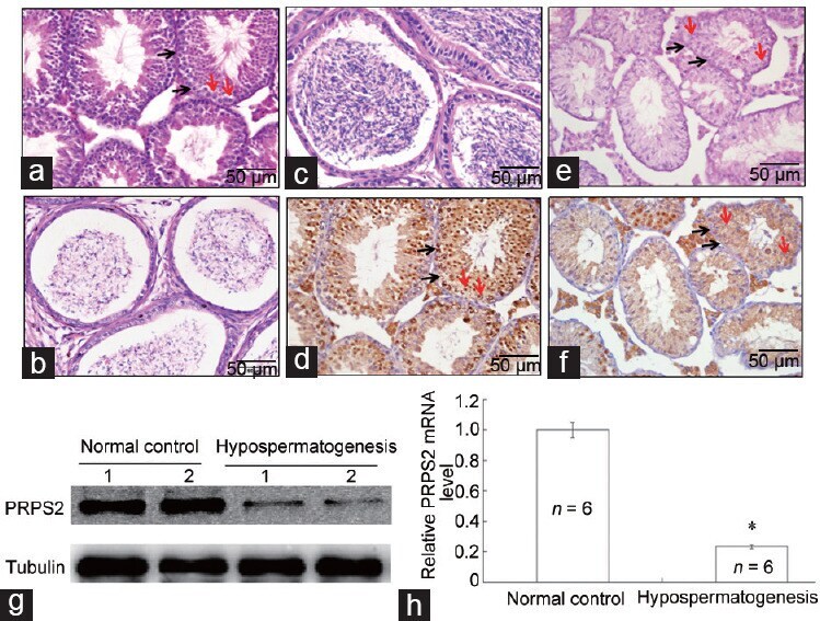
- Figure 2 Correlation of PRPS2 expression with hypospermatogenesis. ( a ) H and E staining of the testis with normal spermatogenesis. ( b ) H and E staining of normal cauda epididymidis. ( c ) H and E staining of a testis with hypospermatogenesis. ( d ) H and E staining of cauda epididymidis in hypospermatogenesis. ( e ) PRPS2 expression was detected in normal testis by IHC. ( f ) PRPS2 expression was detected in a testis with hypospermatogenesis by IHC. Scale bars = 50 mum. ( g ) PRPS2 expression was detected in testis with normal spermatogenesis and hypospermatogenesis by Western blot. ( h ) PRPS2 expression was detected in testis tissues with normal spermatogenesis and hypospermatogenesis by qRT-PCR. *Hypospermatogenesis group compared with normal control, P < 0.05 by independent-samples t -test. Red arrow: spermatogonia; Black arrow: spermatocyte. PRPS2: phosphoribosyl-pyrophosphate synthetase 2; H and E: hematoxylinand and eosin; IHC: immunohistochemical; qRT-PCR: quantitative real-time polymerase chain reaction.
- Submitted by
- Invitrogen Antibodies (provider)
- Main image

- Experimental details
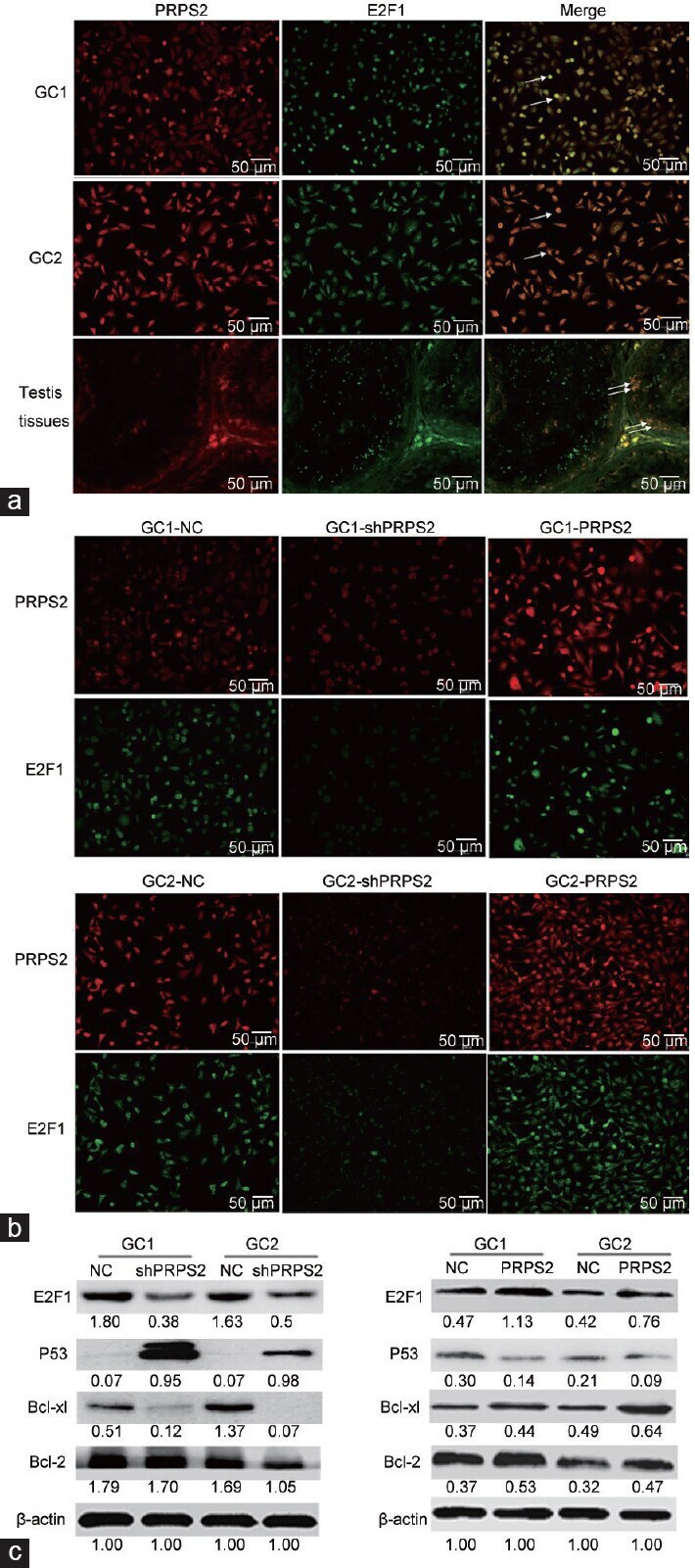
- Figure 6 PRPS2 targetes E2F1 and activation of the P53/Bcl-xl/Bcl-2 signal pathway. ( a ) Colocation of PRPS2 and E2F1 in GC1 and GC2 cells and normal testis tissue was observed by double immunofluorescent staining. ( b ) After transfection, the expression levels of PRPS2 and E2F1 were detected by double immunofluorescent staining. ( c ) Western blot was used to measure the expression levels of the E2F1/P53/Bcl-xl/Bcl-2 signaling pathway. Values beneath the blots are relative to the control. White arrow: colocation. PRPS2: phosphoribosyl-pyrophosphate synthetase 2; NC: negative control; E2F1: E2F transcription factor 1.