 Explore
 Validate
 Learn
Learn Western blot
Antibody data
- Antibody Data
- Antigen structure
- References [0]
- Comments [0]
- Validations
- Western blot [1]
- Immunocytochemistry [2]
- Immunohistochemistry [8]
- Flow cytometry [1]
Submit
Validation data
Reference
Comment
Report error
- Product number
- GTX83534 - Provider product page

- Provider
- GeneTex
- Proper citation
- GeneTex Cat#GTX83534, RRID:AB_10730402
- Product name
- TACC3 antibody [8F6]
- Antibody type
- Monoclonal
- Reactivity
- Human
- Host
- Mouse
No comments: Submit comment
Supportive validation
- Submitted by
- GeneTex (provider)
- Main image
- Experimental details
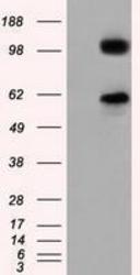
- HEK293T cells were transfected with the pCMV6-ENTRY control (Left lane) or pCMV6-ENTRY TACC3 (Right lane) cDNA for 48 hrs and lysed. Equivalent amounts of cell lysates (5 ug per lane) were separated by SDS-PAGE and immunoblotted with anti-TACC3.
- Validation comment
- WB

Supportive validation
- Submitted by
- GeneTex (provider)
- Main image

- Experimental details
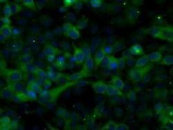
- Anti-TACC3 mouse monoclonal antibody (GTX83534) immunofluorescent staining of COS7 cells transiently transfected with TACC3

- Submitted by
- GeneTex (provider)
- Main image
- Experimental details
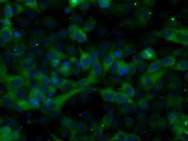
- Immunofluorescent staining of HeLa cells using anti-TACC3 mouse monoclonal antibody (GTX83534).
Supportive validation
- Submitted by
- GeneTex (provider)
- Main image
- Experimental details
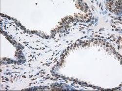
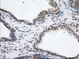
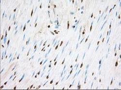
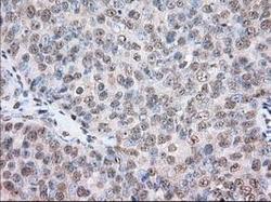
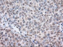
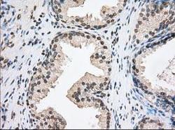
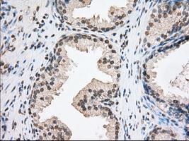
- Immunohistochemical staining of paraffin-embedded Carcinoma of Human prostate tissue using anti-TACC3 mouse monoclonal antibody. (GTX83534, Dilution 1:50)
- Submitted by
- GeneTex (provider)
- Main image
- Experimental details
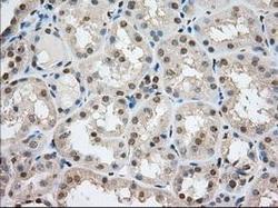
- Immunohistochemical staining of paraffin-embedded Human Kidney tissue using anti-TACC3 mouse monoclonal antibody. (GTX83534, Dilution 1:50)

- Submitted by
- GeneTex (provider)
- Main image
- Experimental details
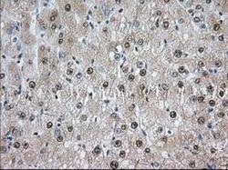
- Immunohistochemical staining of paraffin-embedded Human liver tissue using anti-TACC3 mouse monoclonal antibody. (GTX83534, Dilution 1:50)

- Submitted by
- GeneTex (provider)
- Main image

- Experimental details
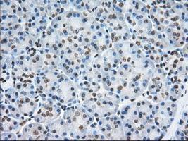
- Immunohistochemical staining of paraffin-embedded Human pancreas tissue using anti-TACC3 mouse monoclonal antibody. (GTX83534, Dilution 1:50)
- Submitted by
- GeneTex (provider)
- Main image
- Experimental details
- Immunohistochemical staining of paraffin-embedded Human colon tissue using anti-TACC3 mouse monoclonal antibody. (GTX83534, Dilution 1:50)

- Submitted by
- GeneTex (provider)
- Main image

- Experimental details
- Immunohistochemical staining of paraffin-embedded Adenocarcinoma of Human colon tissue using anti-TACC3 mouse monoclonal antibody. (GTX83534, Dilution 1:50)

- Submitted by
- GeneTex (provider)
- Main image
- Experimental details
- Immunohistochemical staining of paraffin-embedded Adenocarcinoma of Human ovary tissue using anti-TACC3 mouse monoclonal antibody. (GTX83534, Dilution 1:50)
- Submitted by
- GeneTex (provider)
- Main image
- Experimental details
- Immunohistochemical staining of paraffin-embedded Human prostate tissue using anti-TACC3 mouse monoclonal antibody. (GTX83534, Dilution 1:50)
Supportive validation
- Submitted by
- GeneTex (provider)
- Main image
- Experimental details
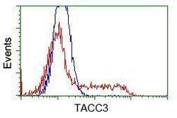
- HEK293T cells transfected with either RC210754 overexpress plasmid(Red) or empty vector control plasmid(Blue) were immunostained by anti-TACC3 antibody(GTX83534), and then analyzed by flow cytometry.
