Explore
 Validate
 Learn
Learn Western blot
Antibody data
- Antibody Data
- Antigen structure
- References [1]
- Comments [0]
- Validations
- Western blot [1]
- Immunohistochemistry [1]
- Flow cytometry [1]
Submit
Validation data
Reference
Comment
Report error
- Product number
- NBP2-24767 - Provider product page

- Provider
- Novus Biologicals
- Product name
- Rabbit Polyclonal TLR7 Antibody
- Antibody type
- Polyclonal
- Description
- Protein G purified.
- Reactivity
- Human, Simian
- Host
- Rabbit
- Isotype
- IgG
- Vial size
- 0.1 mg
- Concentration
- 0.5 mg/ml
- Storage
- Store at 4C short term. Aliquot and store at -20C long term. Avoid freeze-thaw cycles.
Submitted references Increased expression of endosomal members of toll-like receptor family abrogates wound healing in patients with type 2 diabetes mellitus.
Singh K, Agrawal NK, Gupta SK, Mohan G, Chaturvedi S, Singh K
International wound journal 2016 Oct;13(5):927-35
International wound journal 2016 Oct;13(5):927-35
No comments: Submit comment
Supportive validation
- Submitted by
- Novus Biologicals (provider)
- Main image

- Experimental details
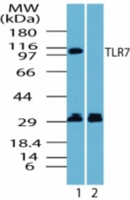
- Western Blot: TLR7 Antibody [NBP2-24767] - Analysis of human TLR7 in Ramos cell lysate in the 1) absence and 2) presence of immunizing peptide using this antibody.
Supportive validation
- Submitted by
- Novus Biologicals (provider)
- Main image
- Experimental details
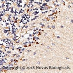
- Immunohistochemistry-Paraffin: TLR7 Antibody [NBP2-24767] - Human brain using 1:50 dilution of TLR7 antibody on a Bond Rx autostainer (Leica Biosystems). The assay involved 20 minutes of heat induced antigen retrieval (HIER) using 10mM sodium citrate buffer (pH 6.0) and endogenous peroxidase quenching with peroxide block. The sections were incubated with primary antibody for 30 minutes and Bond Polymer Refine Detection (Leica Biosystems) with DAB was used for signal development followed by counterstaining with hematoxylin. Whole slide scanning and capturing of representative images (20X) was performed using Aperio AT2 (Leica Biosystems). Punctate staining of neurons was observed. Staining was performed by Histowiz.

Supportive validation
- Submitted by
- Novus Biologicals (provider)
- Main image

- Experimental details
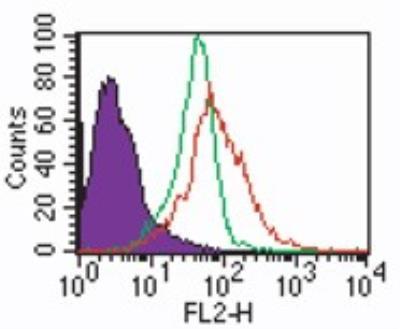
- Flow Cytometry: TLR7 Antibody [NBP2-24767] - Intracellular staining by analysis of TLR7 in human PBMCs using this antibody at 2 ug/ml. Shaded histogrm represents cells alone; green represents rabbit IgG isotype control, and red represents the TLR7 antibody.