Explore
 Validate
 Learn
Learn Western blot
 Immunocytochemistry
Antibody data
- Antibody Data
- Antigen structure
- References [1]
- Comments [0]
- Validations
- Western blot [1]
Submit
Validation data
Reference
Comment
Report error
- Product number
- PB9452 - Provider product page

- Provider
- Boster Biological Technology
- Product name
- Anti-TLR7 Antibody Picoband™
- Antibody type
- Polyclonal
- Description
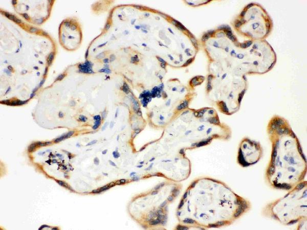
- Polyclonal antibody for TLR7 detection. Host: Rabbit.Size: 100μg/vial. Tested applications: IHC-P. Reactive species: Human. TLR7 information: Molecular Weight: 120922 MW; Subcellular Localization: Endoplasmic reticulum membrane ; Single-pass type I membrane protein . Endosome . Lysosome . Cytoplasmic vesicle, phagosome. Relocalizes from endoplasmic reticulum to endosome and lysosome upon stimulation with agonist; Tissue Specificity: Detected in brain, placenta, spleen, stomach, small intestine, lung and in plasmacytoid pre-dendritic cells.
- Reactivity
- Human, Mouse, Rat
- Host
- Rabbit
- Vial size
- 100μg/vial
- Concentration
- Add 0.2ml of distilled water will yield a concentration of 500ug/ml.
- Storage
- At -20°C for one year. After reconstitution, at 4°C for one month. It can also be aliquoted and stored frozen at -20°C for a longer time. Avoid repeated freezing and thawing.
- Handling
- Add 0.2ml of distilled water will yield a concentration of 500ug/ml.
Submitted references Identification and Analysis of Neutrophil Extracellular Trap-Related Genes in Osteoarthritis by Bioinformatics and Experimental Verification.
Luan T, Yang X, Kuang G, Wang T, He J, Liu Z, Gong X, Wan J, Li K
Journal of inflammation research 2023;16:3837-3852
Journal of inflammation research 2023;16:3837-3852
No comments: Submit comment
Supportive validation
- Submitted by
- Boster Biological Technology (provider)
- Main image
- Experimental details
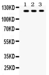
- Western blot analysis of TLR7 using anti-TLR7 antibody (PB9452). Electrophoresis was performed on a 5-20% SDS-PAGE gel at 70V (Stacking gel) / 90V (Resolving gel) for 2-3 hours. The sample well of each lane was loaded with 50ug of sample under reducing conditions. Lane 1: MCF-7 Whole Cell Lysate Lane 2: COLO320 Whole Cell Lysate Lane 3: JURKAT Whole Cell Lysate After Electrophoresis, proteins were transferred to a Nitrocellulose membrane at 150mA for 50-90 minutes. Blocked the membrane with 5% Non-fat Milk/ TBS for 1.5 hour at RT. The membrane was incubated with rabbit anti-TLR7 antigen affinity purified polyclonal antibody (Catalog # PB9452) at 0.5 μg/mL overnight at 4°C, then washed with TBS-0.1%Tween 3 times with 5 minutes each and probed with a goat anti-rabbit IgG-HRP secondary antibody at a dilution of 1:10000 for 1.5 hour at RT. The signal is developed using an Enhanced Chemiluminescent detection (ECL) kit (Catalog # EK1002) with Tanon 5200 system. A specific band was detected for TLR7 at approximately 121KD. The expected band size for TLR7 is at 84KD.
- Additional image