Explore
 Validate
 Learn
Learn Immunohistochemistry
Antibody data
- Antibody Data
- Antigen structure
- References [0]
- Comments [0]
- Validations
- Immunohistochemistry [2]
- Flow cytometry [2]
Submit
Validation data
Reference
Comment
Report error
- Product number
- 10-3015-NALE - Provider product page

- Provider
- ABEOMICS Inc.
- Product name
- Anti-TLR7 Antibody
- Antibody type
- Monoclonal
- Description
- TLR7 (Toll-like receptor 7) is a member of the Toll-like receptor (TLR) family. TLR7 is a nucleotide-sensing TLR which is activated by single-stranded RNA; which plays a fundamental role in pathogen recognition and activation of innate immunity. TLR7 controls host immune response against pathogens through recognition of molecular patterns specific to microorganisims. TLR7 interacts with MYD88 via their respective TIR domains and also interacts with UNC93B1. Post-translational modifications and glycosylation occur in TLR7. TLR7 detected in brain, placenta, spleen, stomach, small intestine, lungs and plasmacytoid pre-dendritic cells.
- Reactivity
- Human
- Host
- Mouse
- Conjugate
- Unconjugated
- Antigen sequence
A partial length recombinant human
TLR7 protein (amino acids 500-900)
was used as the immunogen for this
antibody.- Isotype
- IgG
- Antibody clone number
- ABM2C27
- Vial size
- 100 µg
- Concentration
- 0.5 mg/ml
- Storage
- Store the antibody at 4°C, stable for 6 months. For long-term storage, store at -20°C. Avoid repeat freez thawing
No comments: Submit comment
Supportive validation
- Submitted by
- ABEOMICS Inc. (provider)
- Main image
- Experimental details
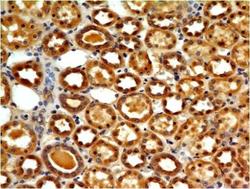
- Immunohistochemical analysis of TLR7 in human Kidney tissue using TLR7 antibody (Clone: ABM2C27) at 5 µg/ml.
- Protocol
- Protocol
- Submitted by
- ABEOMICS Inc. (provider)
- Main image
- Experimental details
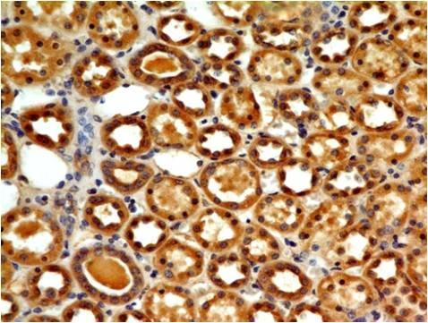
- Immunohistochemical analysis of TLR7 in Renal Cell Carcinoma using TLR7 antibody (Clone: ABM2C27) at 5 µg/ml.
- Protocol
- Protocol

Supportive validation
- Submitted by
- ABEOMICS Inc. (provider)
- Main image
- Experimental details
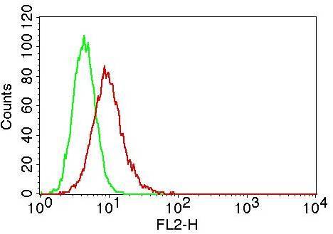
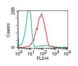
- Intracellular flow analysis of TLR7 in PBMC (Lymphocyte) using 0.5 µg/10^6 cells of TLR7 antibody (Clone: ABM2C27). Green represents isotype control; red represents anti-TLR7 antibody. Goat anti-mouse PE conjugate was used as secondary.
- Protocol
- Protocol
- Submitted by
- ABEOMICS Inc. (provider)
- Main image

- Experimental details
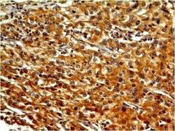
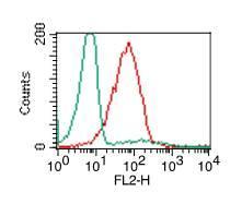
- Intracellular flow analysis of TLR7 in Raji cells using 0.5 µg/10^6 cells of TLR7 antibody (Clone: ABM2C27). Green represents isotype control; red represents anti-TLR7 antibody. Goat anti-mouse PE conjugate was used as secondary antibody.
- Protocol
- Protocol