 Explore
 Validate
 Learn
Learn Western blot
 ELISA
ELISAAntibody data
- Antibody Data
- Antigen structure
- References [0]
- Comments [0]
- Validations
- Western blot [1]
- Immunocytochemistry [1]
- Flow cytometry [2]
Submit
Validation data
Reference
Comment
Report error
- Product number
- NBP1-04349 - Provider product page

- Provider
- Novus Biologicals
- Proper citation
- Novus Cat#NBP1-04349, RRID:AB_1522556
- Product name
- Mouse Monoclonal TLR7 Antibody
- Antibody type
- Monoclonal
- Description
- Protein G purified.
- Reactivity
- Human
- Host
- Mouse
- Isotype
- IgG
- Vial size
- 0.1 ml
- Concentration
- 1.0 mg/ml
- Storage
- Store at 4C short term. Aliquot and store at -20C long term. Avoid freeze-thaw cycles.
No comments: Submit comment
Supportive validation
- Submitted by
- Novus Biologicals (provider)
- Main image

- Experimental details
- Western Blot: TLR7 Antibody (4F4) [NBP1-04349] - The Ramos cell lysates 80ug were resolved by SDS-PAGE, transferred to PVDF membrane and probed with anti-human TLR7 antibody (1:1000). Proteins were visualized using a goat anti-mouse secondary antibody conjugated to HRP and an ECL detection system. Arrow indicates TLR7 protein in human Ramos cells.

Supportive validation
- Submitted by
- Novus Biologicals (provider)
- Main image

- Experimental details
- Immunocytochemistry/Immunofluorescence

Supportive validation
- Submitted by
- Novus Biologicals (provider)
- Main image
- Experimental details
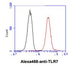
- Flow Cytometry: TLR7 Antibody (4F4) [NBP1-04349] - Analysis of TLR7 in THP-1 cell line, staining at 2-5ug for 1x10⁶cells (red line). The secondary antibody used goat anti-mouse IgG Alexa fluor 488 conjugate. Isotype control antibody was mouse IgG (black line).

- Submitted by
- Novus Biologicals (provider)
- Main image
- Experimental details
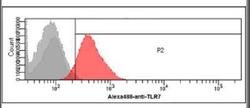
- Flow Cytometry: TLR7 Antibody (4F4) [NBP1-04349] - Flow cytometry analysis of TLR7 in THP-1 cells. The cell was stained with NBP1-04349 at 2-5ug for 1x10⁶cells (red). A Goat anti mouse IgG (Alexa fluor 488) was used as the secondary antibody. Mouse monoclonal IgG was used as the isotype control (dark gray), cells without incubation with primary and secondary antibody was used as the negative control (light gray).
