 Explore
 Validate
 Learn
Learn Western blot
 Immunocytochemistry
Antibody data
- Antibody Data
- Antigen structure
- References [4]
- Comments [0]
- Validations
- Western blot [1]
- Immunohistochemistry [3]
- Flow cytometry [1]
Submit
Validation data
Reference
Comment
Report error
- Product number
- NB100-56682 - Provider product page

- Provider
- Novus Biologicals
- Proper citation
- Novus Cat#NB100-56682, RRID:AB_839015
- Product name
- Rabbit Polyclonal TLR7 Antibody
- Antibody type
- Polyclonal
- Description
- Protein G purified.
- Reactivity
- Human, Mouse
- Host
- Rabbit
- Isotype
- IgG
- Vial size
- 0.1 ml
- Concentration
- 1.0 mg/ml
- Storage
- Store at 4C short term. Aliquot and store at -20C long term. Avoid freeze-thaw cycles.
Submitted references Localization of nucleic acid-sensing toll-like receptors in human and mouse pancreas.
The Expression of Toll-like Receptors in Normal Human and Murine Gastrointestinal Organs and the Effect of Microbiome and Cancer.
Functional effects of Toll-like receptor (TLR)3, 7, 9, RIG-I and MDA-5 stimulation in nasal epithelial cells.
Intracellular signaling mechanisms regulating toll-like receptor-mediated activation of eosinophils.
Helminen O, Huhta H, Kauppila JH, Lehenkari PP, Saarnio J, Karttunen TJ
APMIS : acta pathologica, microbiologica, et immunologica Scandinavica 2017 Feb;125(2):85-92
APMIS : acta pathologica, microbiologica, et immunologica Scandinavica 2017 Feb;125(2):85-92
The Expression of Toll-like Receptors in Normal Human and Murine Gastrointestinal Organs and the Effect of Microbiome and Cancer.
Huhta H, Helminen O, Kauppila JH, Salo T, Porvari K, Saarnio J, Lehenkari PP, Karttunen TJ
The journal of histochemistry and cytochemistry : official journal of the Histochemistry Society 2016 Aug;64(8):470-82
The journal of histochemistry and cytochemistry : official journal of the Histochemistry Society 2016 Aug;64(8):470-82
Functional effects of Toll-like receptor (TLR)3, 7, 9, RIG-I and MDA-5 stimulation in nasal epithelial cells.
Tengroth L, Millrud CR, Kvarnhammar AM, Kumlien Georén S, Latif L, Cardell LO
PloS one 2014;9(6):e98239
PloS one 2014;9(6):e98239
Intracellular signaling mechanisms regulating toll-like receptor-mediated activation of eosinophils.
Wong CK, Cheung PF, Ip WK, Lam CW
American journal of respiratory cell and molecular biology 2007 Jul;37(1):85-96
American journal of respiratory cell and molecular biology 2007 Jul;37(1):85-96
No comments: Submit comment
Supportive validation
- Submitted by
- Novus Biologicals (provider)
- Main image

- Experimental details
- Western Blot: TLR7 Antibody [NB100-56682] - Analysis of TLR7 in Ramos cell lysate using NB100-56682 at 1:500.

Supportive validation
- Submitted by
- Novus Biologicals (provider)
- Main image
- Experimental details
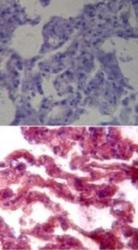
- Immunohistochemistry-Paraffin: TLR7 Antibody [NB100-56682] - Analysis of TLR7 in formalin-fixed, paraffin-embedded human lung tissue using an isotype control (top) and this antibody (bottom) at 1:100.

- Submitted by
- Novus Biologicals (provider)
- Main image
- Experimental details
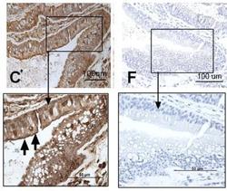
- Immunohistochemistry: TLR7 Antibody [NB100-56682] - Sections of nasal biopsies were incubated with antibodies against TLR9 (C) visualized by 3, 3-diaminobenzidine (brown). In control slides (F), N-series universal negative control reagent was used. All sections were accompanied with a square magnification. All slides were counterstained with haematoxylin (blue). The figure shows one representative biopsy out of four (3 male, 1 female). The arrows indicate positive stained cells. Image collected and cropped by CiteAb from the following publication (//doi.org/10.1371/journal.pone.0098239) licensed under a CC-BY licence.

- Submitted by
- Novus Biologicals (provider)
- Main image
- Experimental details
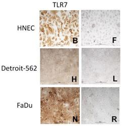
- Immunohistochemistry: TLR7 Antibody [NB100-56682] - Epithelial cells from primary HNEC (B), Detroit-562 (H) and FaDu (N) were incubated with antibody against TLR7 and visualized by 3, 3-diaminobenzidine (brown). In controls, N-series universal negative control reagent was used (F, L, R). All cells were counterstained with haematoxylin (blue). The figure shows one representative staining out of three independent experiments. The markers in the figure are 50 um. The arrows indicate positive stained cells. Image collected and cropped by CiteAb from the following publication (//doi.org/10.1371/journal.pone.0098239) licensed under a CC-BY licence.

Supportive validation
- Submitted by
- Novus Biologicals (provider)
- Main image

- Experimental details
- Flow Cytometry: TLR7 Antibody [NB100-56682] - HNEC, Detroit-562 and FaDu were stained intracellularly with Ab against TLR7 (open histograms) or appropriate isotype control (shaded histograms) and analyzed by flow cytometry. Representative pictures from one out of three independent experiments are shown. Image collected and cropped by CiteAb from the following publication (//doi.org/10.1371/journal.pone.0098239) licensed under a CC-BY licence.
