 Explore
 Validate
 Learn
Learn Western blot
Antibody data
- Antibody Data
- Antigen structure
- References [0]
- Comments [0]
- Validations
- Western blot [3]
- Immunocytochemistry [1]
- Immunohistochemistry [1]
Submit
Validation data
Reference
Comment
Report error
- Product number
- PA5-47875 - Provider product page

- Provider
- Invitrogen Antibodies
- Product name
- TMSB4X Polyclonal Antibody
- Antibody type
- Polyclonal
- Antigen
- Recombinant full-length protein
- Description
- In direct ELISAs, approximately 5% cross-reactivity with recombinant human (rh) Thymosin beta 10 and rhThymosin beta 16 is observed. Reconstitute in sterile PBS to a final concentration of 0.2 mg/mL.
- Reactivity
- Human
- Host
- Sheep
- Isotype
- IgG
- Vial size
- 100 µg
- Concentration
- 0.2 mg/mL
- Storage
- -20° C, Avoid Freeze/Thaw Cycles
No comments: Submit comment
Supportive validation
- Submitted by
- Invitrogen Antibodies (provider)
- Main image

- Experimental details
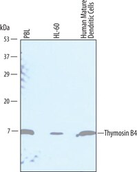
- Western blot analysis from lysates of human peripheral blood lymphocytes (PBL), HL-60 human acute promyelocytic leukemia cell line, and human mature dendritic cells. PVDF Membrane was probed with 1 µg/mL of Sheep Anti-human Thymosin ß4 Antigen Affinity-purified Polyclonal Antibody (Product # PA5-47875) followed by HRP-conjugated Anti-Sheep IgG Secondary Antibody. A specific band was detected for Thymosin ß4 at approximately 5 kDa (as indicated). This experiment was conducted under reducing conditions.

- Submitted by
- Invitrogen Antibodies (provider)
- Main image
- Experimental details
- Western blot analysis of TMSB4X in human peripheral blood lymphocytes (PBL), HL‚60 human acute promyelocytic leukemia cell line, and human mature dendritic cells. Samples were incubated in TMSB4X polyclonal antibody (Product # PA5-47875) using a dilution of 1 µg/mL followed by a HRP-conjugated Anti-Sheep IgG secondary antibody. A specific band was detected for IRF2BP1 at approximately 70 kDa (as indicated). This experiment was conducted under reducing conditions.

- Submitted by
- Invitrogen Antibodies (provider)
- Main image

- Experimental details
- Western blot analysis of TMSB4X in 0.2 mg/mL lysates of HL-60 human acute promyelocytic leukemia cell line. Samples were incubated in TMSB4X polyclonal antibody (Product # PA5-47875) using a dilution of 10 µg/mL followed by HRP-conjugated Anti-Sheep IgG at a dilution of 0.0763888888888889. A specific band was detected for Thymosin β4 at approximately 8 kDa (as indicated). This experiment was conducted under reducing conditions and using the 2-40 kDa separation system.

Supportive validation
- Submitted by
- Invitrogen Antibodies (provider)
- Main image

- Experimental details
- Immunocytochemistry analysis of TMSB4X in immersion fixed HeLa human cervical epithelial carcinoma cell line. Samples were incubated in TMSB4X polyclonal antibody (Product # PA5-47875) using a dilution of 10 µg/mL for 3 hours at room temperature followed by NorthernLights™ 557-conjugated Anti-Sheep IgG Secondary Antibody (red, upper panel) and counterstained with DAPI (blue, lower panel). Specific staining was localized to cytoplasm.

Supportive validation
- Submitted by
- Invitrogen Antibodies (provider)
- Main image

- Experimental details
- Immunocytochemical analysis of Thymosin ß4 was detected in immersion fixed HeLa human cervical epithelial carcinoma cell line using Sheep Anti-human Thymosin ß4 Antigen Affinity-purified Polyclonal Antibody (Product # PA5-47875) at 10 µg/mL for 3 hours at room temperature. Cells were stained using the 557-conjugated Anti-Sheep IgG Secondary Antibody (red, upper pane and counterstained with DAPI (blue, lower panel). Specific staining was localized to cytoplasm.
