 Explore
 Validate
 Learn
Learn Western blot
 Immunohistochemistry
Antibody data
- Antibody Data
- Antigen structure
- References [16]
- Comments [0]
- Validations
- Immunohistochemistry [1]
Submit
Validation data
Reference
Comment
Report error
- Product number
- HPA001888 - Provider product page

- Provider
- Atlas Antibodies
- Proper citation
- Atlas Antibodies Cat#HPA001888, RRID:AB_1079276
- Product name
- Anti-LRG1
- Antibody type
- Polyclonal
- Description
- Polyclonal Antibody against Human LRG1, Gene description: leucine-rich alpha-2-glycoprotein 1, Alternative Gene Names: LRG, Validated applications: IHC, WB, Uniprot ID: P02750, Storage: Store at +4°C for short term storage. Long time storage is recommended at -20°C.
- Reactivity
- Human
- Host
- Rabbit
- Conjugate
- Unconjugated
- Isotype
- IgG
- Vial size
- 100 µl
- Concentration
- 0.2 mg/ml
- Storage
- Store at +4°C for short term storage. Long time storage is recommended at -20°C.
- Handling
- The antibody solution should be gently mixed before use.
Submitted references LRG1 and SDR16C5 protein expressions differ according to HPV status in oropharyngeal squamous cell carcinoma.
Crystal structure of LRG1 and the functional significance of LRG1 glycan for LPHN2 activation
Latrophilin-2 is a novel receptor of LRG1 that rescues vascular and neurological abnormalities and restores diabetic erectile function
LRG1 is an adipokine that promotes insulin sensitivity and suppresses inflammation
Novel Blood Vascular Endothelial Subtype-Specific Markers in Human Skin Unearthed by Single-Cell Transcriptomic Profiling.
LRG1 Expression Is Elevated in the Eyes of Patients with Neovascular Age-Related Macular Degeneration
Evaluation of leucine-rich alpha-2 glycoprotein as a biomarker of fetal infection.
Leucine-rich alpha 2 glycoprotein is a new marker for active disease of tuberculosis.
A High-throughput Bead-based Affinity Assay Enables Analysis of Genital Protein Signatures in Women At Risk of HIV Infection
Leucine-rich alpha-2-glycoprotein-1 is up-regulated in colorectal cancer and is a tumor promoter
LRG1 promotes proliferation and inhibits apoptosis in colorectal cancer cells via RUNX1 activation
Overexpression of leucine-rich α2-glycoprotein-1 is a prognostic marker and enhances tumor migration in gastric cancer.
Sputum Leucine-Rich Alpha-2 Glycoprotein as a Marker of Airway Inflammation in Asthma.
Analysis of the Cerebrospinal Fluid Proteome in Alzheimer's Disease.
LRG1 promotes angiogenesis by modulating endothelial TGF-β signalling
Tumour expression of bladder cancer‐associated urinary proteins
Randén-Brady R, Carpén T, Hautala LC, Tolvanen T, Haglund C, Joenväärä S, Mattila P, Mäkitie A, Lehtonen S, Hagström J, Silén S
Scientific reports 2024 Jun 19;14(1):14148
Scientific reports 2024 Jun 19;14(1):14148
Crystal structure of LRG1 and the functional significance of LRG1 glycan for LPHN2 activation
Yang J, Yin G, Kim D, Han A, Lee D, Min K, Fu Y, Yun J, Suh J, Ryu J, Kim H
Experimental & Molecular Medicine 2023;55(5):1013-1022
Experimental & Molecular Medicine 2023;55(5):1013-1022
Latrophilin-2 is a novel receptor of LRG1 that rescues vascular and neurological abnormalities and restores diabetic erectile function
Yin G, Kim D, Kang J, Im Y, Lee D, Han A, Ock J, Choi M, Kwon M, Limanjaya A, Jung S, Yang J, Min K, Yun J, Koh Y, Park J, Hwang D, Suh J, Ryu J, Kim H
Experimental & Molecular Medicine 2022;54(5):626-638
Experimental & Molecular Medicine 2022;54(5):626-638
LRG1 is an adipokine that promotes insulin sensitivity and suppresses inflammation
Choi C, Barr W, Zaman S, Model C, Park A, Koenen M, Lin Z, Szwed S, Marchildon F, Crane A, Carroll T, Molina H, Cohen P
eLife 2022;11
eLife 2022;11
Novel Blood Vascular Endothelial Subtype-Specific Markers in Human Skin Unearthed by Single-Cell Transcriptomic Profiling.
He Y, Tacconi C, Dieterich LC, Kim J, Restivo G, Gousopoulos E, Lindenblatt N, Levesque MP, Claassen M, Detmar M
Cells 2022 Mar 25;11(7)
Cells 2022 Mar 25;11(7)
LRG1 Expression Is Elevated in the Eyes of Patients with Neovascular Age-Related Macular Degeneration
Mundo L, Tosi G, Lazzi S, Pertile G, Parolini B, Neri G, Posarelli M, De Benedetto E, Bacci T, Silvestri E, Siciliano M, Barbera S, Orlandini M, Greenwood J, Moss S, Galvagni F
International Journal of Molecular Sciences 2021;22(16):8879
International Journal of Molecular Sciences 2021;22(16):8879
Evaluation of leucine-rich alpha-2 glycoprotein as a biomarker of fetal infection.
Kajimoto E, Endo M, Fujimoto M, Matsuzaki S, Fujii M, Yagi K, Kakigano A, Mimura K, Tomimatsu T, Serada S, Takeuchi M, Yoshino K, Ueda Y, Kimura T, Naka T
PloS one 2020;15(11):e0242076
PloS one 2020;15(11):e0242076
Leucine-rich alpha 2 glycoprotein is a new marker for active disease of tuberculosis.
Fujimoto M, Matsumoto T, Serada S, Tsujimura Y, Hashimoto S, Yasutomi Y, Naka T
Scientific reports 2020 Feb 25;10(1):3384
Scientific reports 2020 Feb 25;10(1):3384
A High-throughput Bead-based Affinity Assay Enables Analysis of Genital Protein Signatures in Women At Risk of HIV Infection
Månberg A, Bradley F, Qundos U, Guthrie B, Birse K, Noël-Romas L, Lindskog C, Bosire R, Kiarie J, Farquhar C, Burgener A, Nilsson P, Broliden K
Molecular & Cellular Proteomics 2019;18(3):461-476
Molecular & Cellular Proteomics 2019;18(3):461-476
Leucine-rich alpha-2-glycoprotein-1 is up-regulated in colorectal cancer and is a tumor promoter
Zhang Q, Huang R, Tang Q, Yu Y, Huang Q, Chen Y, Wang G, Wang X
OncoTargets and Therapy 2018;Volume 11
OncoTargets and Therapy 2018;Volume 11
LRG1 promotes proliferation and inhibits apoptosis in colorectal cancer cells via RUNX1 activation
Ahmad A, Zhou Y, Zhang X, Zhang J, Fang J, Ge Z, Li X
PLOS ONE 2017;12(4):e0175122
PLOS ONE 2017;12(4):e0175122
Overexpression of leucine-rich α2-glycoprotein-1 is a prognostic marker and enhances tumor migration in gastric cancer.
Yamamoto M, Takahashi T, Serada S, Sugase T, Tanaka K, Miyazaki Y, Makino T, Kurokawa Y, Yamasaki M, Nakajima K, Takiguchi S, Naka T, Mori M, Doki Y
Cancer science 2017 Oct;108(10):2052-2060
Cancer science 2017 Oct;108(10):2052-2060
Sputum Leucine-Rich Alpha-2 Glycoprotein as a Marker of Airway Inflammation in Asthma.
Honda H, Fujimoto M, Miyamoto S, Ishikawa N, Serada S, Hattori N, Nomura S, Kohno N, Yokoyama A, Naka T
PloS one 2016;11(9):e0162672
PloS one 2016;11(9):e0162672
Analysis of the Cerebrospinal Fluid Proteome in Alzheimer's Disease.
Khoonsari PE, Häggmark A, Lönnberg M, Mikus M, Kilander L, Lannfelt L, Bergquist J, Ingelsson M, Nilsson P, Kultima K, Shevchenko G
PloS one 2016;11(3):e0150672
PloS one 2016;11(3):e0150672
LRG1 promotes angiogenesis by modulating endothelial TGF-β signalling
Wang X, Abraham S, McKenzie J, Jeffs N, Swire M, Tripathi V, Luhmann U, Lange C, Zhai Z, Arthur H, Bainbridge J, Moss S, Greenwood J
Nature 2013;499(7458):306-311
Nature 2013;499(7458):306-311
Tumour expression of bladder cancer‐associated urinary proteins
Lindén M, Segersten U, Runeson M, Wester K, Busch C, Pettersson U, Lind S, Malmström P
BJU International 2013;112(3):407-415
BJU International 2013;112(3):407-415
No comments: Submit comment
Supportive validation
- Submitted by
- Atlas Antibodies (provider)
- Enhanced method
- Orthogonal validation
- Main image
- Experimental details
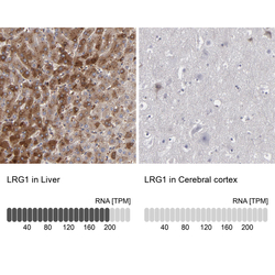
- Immunohistochemistry analysis in human liver and cerebral cortex tissues using HPA001888 antibody. Corresponding LRG1 RNA-seq data are presented for the same tissues.
- Sample type
- Human
- Protocol
- Protocol
