Explore
 Validate
 Learn
Learn Western blot
Antibody data
- Antibody Data
- Antigen structure
- References [1]
- Comments [0]
- Validations
- Western blot [2]
- Immunohistochemistry [1]
Submit
Validation data
Reference
Comment
Report error
- Product number
- AF5407 - Provider product page

- Provider
- Novus Biologicals
- Product name
- Sheep Polyclonal DYRK1A Antibody
- Antibody type
- Polyclonal
- Description
- Immunogen affinity purified. Detects human and rat DYRK1A in Western blots.
- Reactivity
- Human, Rat
- Host
- Sheep
- Conjugate
- Unconjugated
- Isotype
- IgG
- Vial size
- 100 ug
- Concentration
- LYOPH
- Storage
- Use a manual defrost freezer and avoid repeated freeze-thaw cycles. 12 months from date of receipt, -20 to -70 degreesC as supplied. 1 month, 2 to 8 degreesC under sterile conditions after reconstitution. 6 months, -20 to -70 degreesC under sterile conditions after reconstitution.
Submitted references Truncation and Activation of Dual Specificity Tyrosine Phosphorylation-regulated Kinase 1A by Calpain I: A MOLECULAR MECHANISM LINKED TO TAU PATHOLOGY IN ALZHEIMER DISEASE.
Jin N, Yin X, Gu J, Zhang X, Shi J, Qian W, Ji Y, Cao M, Gu X, Ding F, Iqbal K, Gong CX, Liu F
The Journal of biological chemistry 2015 Jun 12;290(24):15219-37
The Journal of biological chemistry 2015 Jun 12;290(24):15219-37
No comments: Submit comment
Supportive validation
- Submitted by
- Novus Biologicals (provider)
- Main image
- Experimental details
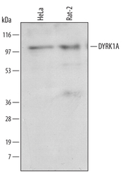
- Detection of Human/Rat DYRK1A by Western Blot. Western blot shows lysates of HeLa human cervical epithelial carcinoma cell line and Rat-2 rat embryonic fibroblast cell line. PVDF membrane was probed with 1 µg/mL of Sheep Anti-Human/Rat DYRK1A Antigen Affinity-purified Polyclonal Antibody (Catalog # AF5407) followed by HRP-conjugated Anti-Sheep IgG Secondary Antibody (Catalog # HAF016). A specific band was detected for DYRK1A at approximately 100 kDa (as indicated). This experiment was conducted under reducing conditions and using Immunoblot Buffer Group 1.

- Submitted by
- Novus Biologicals (provider)
- Main image
- Experimental details
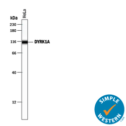
- Detection of Human DYRK1A by Simple WesternTM. Simple Western lane view shows lysates of HeLa human cervical epithelial carcinoma cell line, loaded at 0.2 mg/mL. A specific band was detected for DYRK1A at approximately 113 kDa (as indicated) using 10 µg/mL of Sheep Anti-Human/Rat DYRK1A Antigen Affinity-purified Polyclonal Antibody (Catalog # AF5407) followed by 1:50 dilution of HRP-conjugated Anti-Sheep IgG Secondary Antibody (Catalog # HAF016) . This experiment was conducted under reducing conditions and using the 12-230 kDa separation system.

Supportive validation
- Submitted by
- Novus Biologicals (provider)
- Main image

- Experimental details
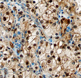
- DYRK1A in Human Kidney. DYRK1A was detected in immersion fixed paraffin-embedded sections of human kidney using Sheep Anti-Human/Rat DYRK1A Antigen Affinity-purified Polyclonal Antibody (Catalog # AF5407) at 10 µg/mL overnight at 4 °C. Before incubation with the primary antibody, tissue was subjected to heat-induced epitope retrieval using Antigen Retrieval Reagent-Basic (Catalog # CTS013). Tissue was stained using the Anti-Sheep HRP-DAB Cell & Tissue Staining Kit (brown; Catalog # CTS019) and counterstained with hematoxylin (blue). Specific staining was localized to nuclei of epithelial cells in convoluted tubules. View our protocol for Chromogenic IHC Staining of Paraffin-embedded Tissue Sections. This application has not been tested in rat samples.