Explore
 Validate
 Learn
Learn Western blot
 Immunocytochemistry
Antibody data
- Antibody Data
- Antigen structure
- References [1]
- Comments [0]
- Validations
- Immunocytochemistry [2]
- Immunohistochemistry [6]
- Other assay [1]
Submit
Validation data
Reference
Comment
Report error
- Product number
- MA5-24621 - Provider product page

- Provider
- Invitrogen Antibodies
- Product name
- NSD2 Monoclonal Antibody (CL1063)
- Antibody type
- Monoclonal
- Antigen
- Recombinant full-length protein
- Description
- Immunogen sequence: SANGKTPSCE VNRECSVFLS KAQLSSSLQE GVMQKFNGHD ALPFIPADKL KDLTSRVFNG EPGAHDAKLR FESQEMKGIG TPPNTTPIKN GSPEIKLKIT KTYMNGKPLF ESSICGD Highest antigen sequence identity to the following orthologs: Mouse - 91%, Rat - 91%. Binds to an epitope located within the peptide sequence ALPFIPADKL as determined by overlapping synthetic peptides.
- Reactivity
- Human
- Host
- Mouse
- Isotype
- IgG
- Antibody clone number
- CL1063
- Vial size
- 100 μL
- Concentration
- 1 mg/mL
- Storage
- Store at 4°C short term. For long term storage, store at -20°C, avoiding freeze/thaw cycles.
Submitted references Activating transcription factor 2 promotes the progression of hepatocellular carcinoma by inducing the activation of the WHSC1-mediated TOP2A/PI3K/AKT axis.
Bao ZM, Yao D, Qian X, Zhang HG, Yang M, Guo YH, Qin L
The Kaohsiung journal of medical sciences 2022 Jul;38(7):662-674
The Kaohsiung journal of medical sciences 2022 Jul;38(7):662-674
No comments: Submit comment
Supportive validation
- Submitted by
- Invitrogen Antibodies (provider)
- Main image

- Experimental details
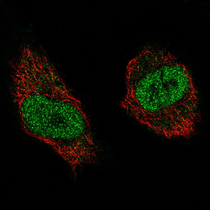
- Immunocytochemistry-Immunofluorescence analysis of NSD2 in U-251 cells using NSD2 Monoclonal Antibody (CL1063) (Product # MA5-24621), showing specific staining in the nucleoplasm in green. Microtubule- and nuclear probes are visualized in red and blue, respectively (where available).

- Submitted by
- Invitrogen Antibodies (provider)
- Main image

- Experimental details
- Immunofluorecent analysis of NSD2 in U-251 cells using NSD2 Monoclonal Antibody (CL1063) (Product # MA5-24621). Staining shows specific staining in the nucleoplasm in green. Microtubule- and nuclear probes are visualized in red and blue, respectively (where available).
Supportive validation
- Submitted by
- Invitrogen Antibodies (provider)
- Main image
- Experimental details
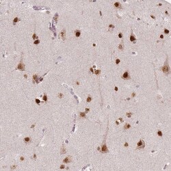
- Immunohistochemical analysis of NSD2 in human cerebral cortex using NSD2 Monoclonal Antibody (CL1063) (Product # MA5-24621) shows strong nuclear positivity in neurons.

- Submitted by
- Invitrogen Antibodies (provider)
- Main image
- Experimental details
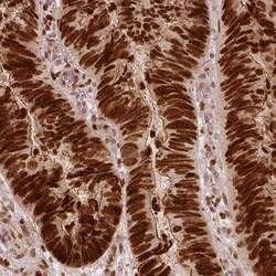
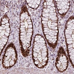
- Immunohistochemical analysis of NSD2 in human colorectal cancer using NSD2 Monoclonal Antibody (CL1063) (Product # MA5-24621) shows strong nuclear positivity in tumor cells.

- Submitted by
- Invitrogen Antibodies (provider)
- Main image
- Experimental details
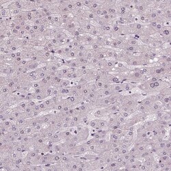
- Immunohistochemical analysis of NSD2 in human liver using NSD2 Monoclonal Antibody (CL1063) (Product # MA5-24621) shows no positivity in hepatocytes as expected.

- Submitted by
- Invitrogen Antibodies (provider)
- Main image
- Experimental details
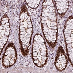
- Immunohistochemical analysis of NSD2 in human rectum using NSD2 Monoclonal Antibody (CL1063) (Product # MA5-24621) shows strong nuclear positivity in glandular cells.

- Submitted by
- Invitrogen Antibodies (provider)
- Main image
- Experimental details
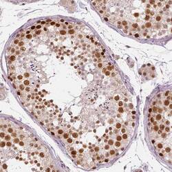
- Immunohistochemical analysis of NSD2 in human testis using NSD2 Monoclonal Antibody (CL1063) (Product # MA5-24621) shows moderate to strong nuclear positivity in cells in seminiferous ducts.

- Submitted by
- Invitrogen Antibodies (provider)
- Main image
- Experimental details
- Immunohistochemical analysis of NSD2 in human rectum using NSD2 Monoclonal Antibody (CL1063) (Product # MA5-24621) shows strong nuclear positivity in glandular cells.

Supportive validation
- Submitted by
- Invitrogen Antibodies (provider)
- Main image
- Experimental details
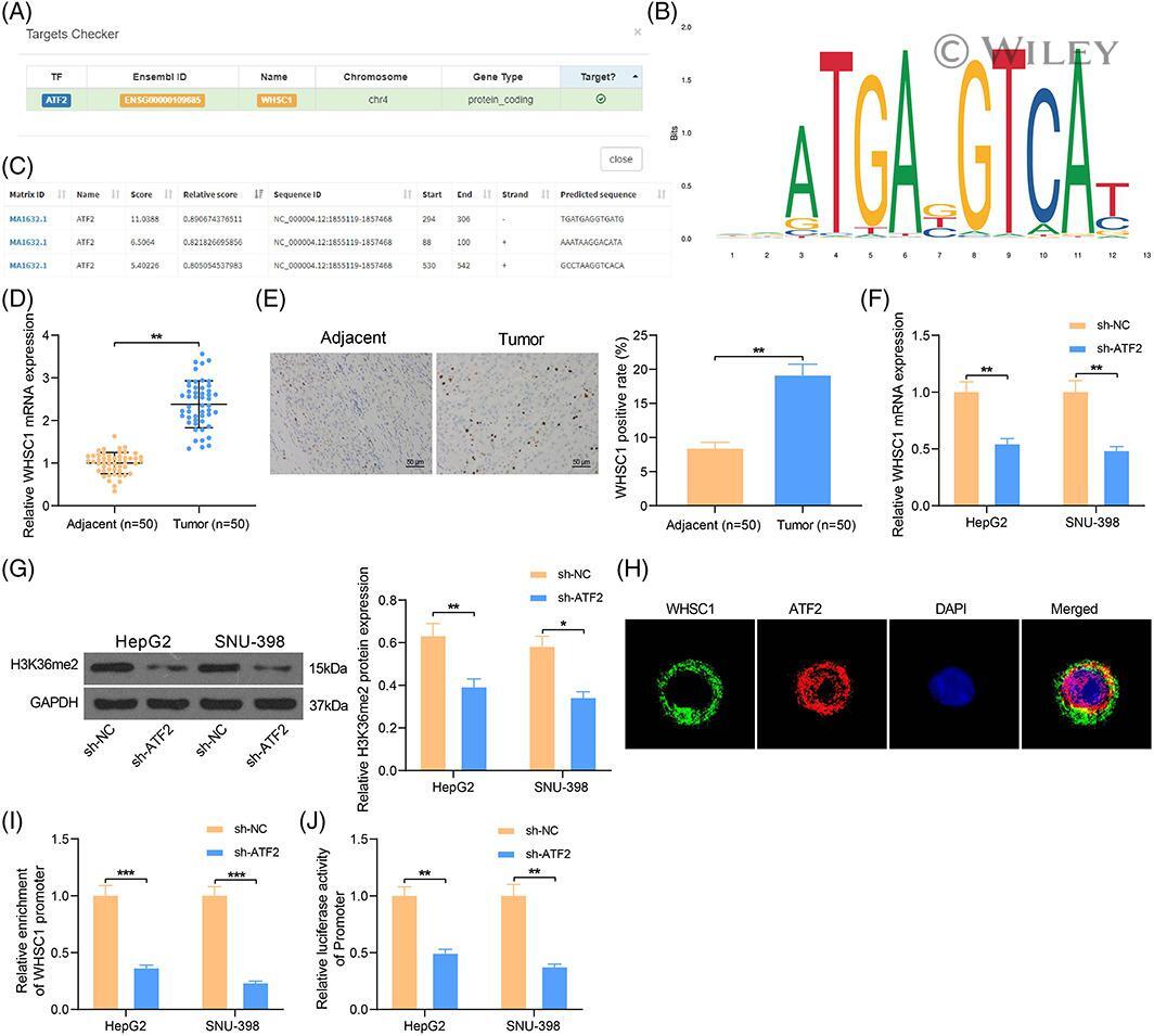
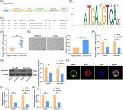
- 3 FIGURE ATF2 silencing reduces expression of WHSC1 in HCC cells. (A) data in the hTFtarget database suggests WHSC1 as a downstream target of ATF2; (B) conservative binding site of ATF2; (C) putative binding site between WHSC1 and ATF2; (D,E) mRNA (D) and protein (E) expression of WHSC1in clinically collected HCC tumor tissues and the paired normal tissues ( n = 50) examined by RT-qPCR and IHC staining (paired t test); (F) mRNA expression of WHSC1 in HepG2 and SNU-398 cells after ATF2 silencing examined by RT-qPCR (two-way ANOVA); (G) protein level of H3K36me2 in HepG2 and SNU-398 cells after ATF2 silencing determined by western blot analysis (two-way ANOVA); (H), distribution of WHSC1 and H3K36me2 in HepG2 cells examined by fluorescence co-localization assay; (I), abundance of WHSC1 promoter fragments enriched by anti-ATF2 determined by the ChIP-qPCR assay (two-way ANOVA); (J), binding relationship between WHSC1 promoter and ATF2 validated by a dual-luciferase reporter gene assay (two-way ANOVA). Data are expressed as the mean +- SD. Repetition = 3. * p < 0.05; ** p < 0.01; *** p < 0.001