Antibody data
- Antibody Data
- Antigen structure
- References [8]
- Comments [0]
- Validations
- Western blot [3]
- Immunohistochemistry [1]
Submit
Validation data
Reference
Comment
Report error
- Product number
- AF1177 - Provider product page

- Provider
- Novus Biologicals
- Product name
- Goat Polyclonal Serpin F1/PEDF Antibody
- Antibody type
- Polyclonal
- Description
- Antigen Affinity-purified. Detects human Serpin F1/PEDF in direct ELISAs. Detects human and mouse Serpin F1/PEDF in Western blots.
- Reactivity
- Human, Mouse
- Host
- Goat
- Conjugate
- Unconjugated
- Isotype
- IgG
- Vial size
- 100 ug
- Concentration
- LYOPH
- Storage
- Use a manual defrost freezer and avoid repeated freeze-thaw cycles. 12 months from date of receipt, -20 to -70 degreesC as supplied. 1 month, 2 to 8 degreesC under sterile conditions after reconstitution. 6 months, -20 to -70 degreesC under sterile conditions after reconstitution.
Submitted references Pigment epithelium-derived factor mediates retinal ganglion cell neuroprotection by suppression of caspase-2.
HtrA1 Mediated Intracellular Effects on Tubulin Using a Polarized RPE Disease Model.
Quantitative changes in the protein and miRNA cargo of plasma exosome-like vesicles after exposure to ionizing radiation.
Secretome and degradome profiling shows that Kallikrein-related peptidases 4, 5, 6, and 7 induce TGFβ-1 signaling in ovarian cancer cells.
Pigment epithelium-derived factor (PEDF) binds to caveolin-1 and inhibits the pro-inflammatory effects of caveolin-1 in endothelial cells.
Long-term retinal PEDF overexpression prevents neovascularization in a murine adult model of retinopathy.
A therapeutic strategy for choroidal neovascularization based on recruitment of mesenchymal stem cells to the sites of lesions.
Inhibition of nuclear translocation of apoptosis-inducing factor is an essential mechanism of the neuroprotective activity of pigment epithelium-derived factor in a rat model of retinal degeneration.
Vigneswara V, Ahmed Z
Cell death & disease 2019 Feb 4;10(2):102
Cell death & disease 2019 Feb 4;10(2):102
HtrA1 Mediated Intracellular Effects on Tubulin Using a Polarized RPE Disease Model.
Melo E, Oertle P, Trepp C, Meistermann H, Burgoyne T, Sborgi L, Cabrera AC, Chen CY, Hoflack JC, Kam-Thong T, Schmucki R, Badi L, Flint N, Ghiani ZE, Delobel F, Stucki C, Gromo G, Einhaus A, Hornsperger B, Golling S, Siebourg-Polster J, Gerber F, Bohrmann B, Futter C, Dunkley T, Hiller S, Schilling O, Enzmann V, Fauser S, Plodinec M, Iacone R
EBioMedicine 2018 Jan;27:258-274
EBioMedicine 2018 Jan;27:258-274
Quantitative changes in the protein and miRNA cargo of plasma exosome-like vesicles after exposure to ionizing radiation.
Yentrapalli R, Merl-Pham J, Azimzadeh O, Mutschelknaus L, Peters C, Hauck SM, Atkinson MJ, Tapio S, Moertl S
International journal of radiation biology 2017 Jun;93(6):569-580
International journal of radiation biology 2017 Jun;93(6):569-580
Secretome and degradome profiling shows that Kallikrein-related peptidases 4, 5, 6, and 7 induce TGFβ-1 signaling in ovarian cancer cells.
Shahinian H, Loessner D, Biniossek ML, Kizhakkedathu JN, Clements JA, Magdolen V, Schilling O
Molecular oncology 2014 Feb;8(1):68-82
Molecular oncology 2014 Feb;8(1):68-82
Pigment epithelium-derived factor (PEDF) binds to caveolin-1 and inhibits the pro-inflammatory effects of caveolin-1 in endothelial cells.
Matsui T, Higashimoto Y, Taira J, Yamagishi S
Biochemical and biophysical research communications 2013 Nov 15;441(2):405-10
Biochemical and biophysical research communications 2013 Nov 15;441(2):405-10
Long-term retinal PEDF overexpression prevents neovascularization in a murine adult model of retinopathy.
Haurigot V, Villacampa P, Ribera A, Bosch A, Ramos D, Ruberte J, Bosch F
PloS one 2012;7(7):e41511
PloS one 2012;7(7):e41511
A therapeutic strategy for choroidal neovascularization based on recruitment of mesenchymal stem cells to the sites of lesions.
Hou HY, Liang HL, Wang YS, Zhang ZX, Wang BR, Shi YY, Dong X, Cai Y
Molecular therapy : the journal of the American Society of Gene Therapy 2010 Oct;18(10):1837-45
Molecular therapy : the journal of the American Society of Gene Therapy 2010 Oct;18(10):1837-45
Inhibition of nuclear translocation of apoptosis-inducing factor is an essential mechanism of the neuroprotective activity of pigment epithelium-derived factor in a rat model of retinal degeneration.
Murakami Y, Ikeda Y, Yonemitsu Y, Onimaru M, Nakagawa K, Kohno R, Miyazaki M, Hisatomi T, Nakamura M, Yabe T, Hasegawa M, Ishibashi T, Sueishi K
The American journal of pathology 2008 Nov;173(5):1326-38
The American journal of pathology 2008 Nov;173(5):1326-38
No comments: Submit comment
Supportive validation
- Submitted by
- Novus Biologicals (provider)
- Main image

- Experimental details
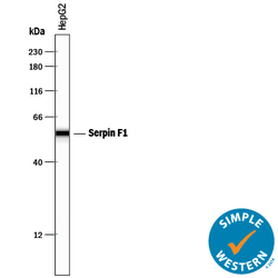
- Detection of Human Serpin F1/PEDF by Simple WesternTM. Simple Western lane view shows lysates of HepG2 human hepatocellular carcinoma cell line, loaded at 0.2 mg/mL. A specific band was detected for Serpin F1/PEDF at approximately 56 kDa (as indicated) using 2 µg/mL of Goat Anti-Human/Mouse Serpin F1/PEDF Antigen Affinity-purified Polyclonal Antibody (Catalog # AF1177) followed by 1:50 dilution of HRP-conjugated Anti-Goat IgG Secondary Antibody (Catalog # HAF109). This experiment was conducted under reducing conditions and using the 12-230 kDa separation system.
- Submitted by
- Novus Biologicals (provider)
- Main image

- Experimental details
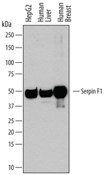
- Detection of Human Serpin F1/PEDF by Western Blot. Western blot shows lysates of HepG2 human hepatocellular carcinoma cell line, human liver tissue, and human breast tissue. PVDF membrane was probed with 0.2 µg/mL of Goat Anti-Human/Mouse Serpin F1/PEDF Antigen Affinity-purified Polyclonal Antibody (Catalog # AF1177) followed by HRP-conjugated Anti-Goat IgG Secondary Antibody (Catalog # HAF109). A specific band was detected for Serpin F1/PEDF at approximately 50 kDa (as indicated). This experiment was conducted under reducing conditions and using Immunoblot Buffer Group 1.
- Submitted by
- Novus Biologicals (provider)
- Main image

- Experimental details
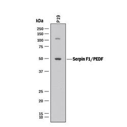
- Detection of Mouse Serpin F1/PEDF by Western Blot. Western blot shows lysates of P19 mouse embryonal carcinoma cell line. PVDF membrane was probed with 2 µg/mL of Goat Anti-Human/Mouse Serpin F1/PEDF Antigen Affinity-purified Polyclonal Antibody (Catalog # AF1177) followed by HRP-conjugated Anti-Goat IgG Secondary Antibody (Catalog # HAF017). A specific band was detected for Serpin F1/PEDF at approximately 50 kDa (as indicated). This experiment was conducted under reducing conditions and using Immunoblot Buffer Group 1.
Supportive validation
- Submitted by
- Novus Biologicals (provider)
- Main image

- Experimental details
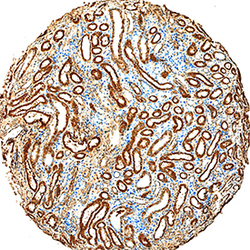
- Serpin F1/PEDF in Human Kidney. Serpin F1/PEDF was detected in immersion fixed paraffin-embedded sections of human kidney using Goat Anti-Human/Mouse Serpin F1/PEDF Antigen Affinity-purified Polyclonal Antibody (Catalog # AF1177) at 15 µg/mL overnight at 4 °C. Tissue was stained using the Anti-Goat HRP-DAB Cell & Tissue Staining Kit (brown; Catalog # CTS008) and counterstained with hematoxylin (blue). Specific staining was localized to convoluted tubules. View our protocol for Chromogenic IHC Staining of Paraffin-embedded Tissue Sections.
 Explore
 Validate
 Learn
Learn Western blot