 Explore
 Validate
 Learn
Learn Western blot
 ELISA
ELISAAntibody data
- Antibody Data
- Antigen structure
- References [0]
- Comments [0]
- Validations
- Western blot [2]
- Immunocytochemistry [4]
- Immunoprecipitation [1]
- Flow cytometry [2]
- Other assay [1]
Submit
Validation data
Reference
Comment
Report error
- Product number
- MA5-29009 - Provider product page

- Provider
- Invitrogen Antibodies
- Product name
- Angiotensinogen Recombinant Rabbit Monoclonal Antibody (1)
- Antibody type
- Monoclonal
- Antigen
- Recombinant full-length protein
- Description
- This product is preservative free. It is recommended to add sodium azide to avoid contamination (final concentration 0.05%-0.1%). Recombinant rabbit monoclonal antibodies are produced using in vitro expression systems. The expression systems are developed by cloning in the specific antibody DNA sequences from immunoreactive rabbits. Then, individual clones are screened to select the best candidates for production. The advantages of using recombinant rabbit monoclonal antibodies include: better specificity and sensitivity, lot-to-lot consistency, animal origin-free formulations, and broader immunoreactivity to diverse targets due to larger rabbit immune repertoire. This antibody has specificity for Human SerpinA8/Angiotensinogen.
- Reactivity
- Human
- Host
- Rabbit
- Isotype
- IgG
- Antibody clone number
- 1
- Vial size
- 100 μL
- Concentration
- 1 mg/mL
- Storage
- Store at 4°C short term. For long term storage, store at -20°C, avoiding freeze/thaw cycles.
No comments: Submit comment
Supportive validation
- Submitted by
- Invitrogen Antibodies (provider)
- Main image
- Experimental details
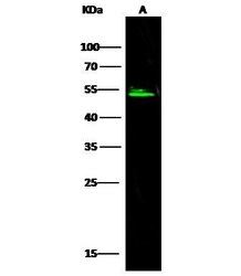
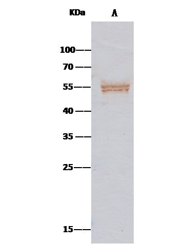
- Western Blot using Angiotensinogen Recombinant Rabbit Monoclonal Antibody (1) (Product # MA5-29009) at 1:500 dilution. Lane A: HepG2 Whole Cell Lysate. Lysates/proteins at 30 μg per lane. Secondary Goat Anti-Rabbit IgG H&L (DyLight™ 800) at 1:10,000 dilution. Developed using the Odyssey technique. Performed under reducing conditions. Predicted band size: 53 kDa. Observed band size: 53 kDa.

- Submitted by
- Invitrogen Antibodies (provider)
- Main image
- Experimental details
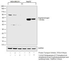
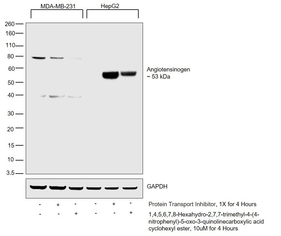
- Western blot was performed using Anti-Angiotensinogen Rabbit Monoclonal Antibody (Product # MA5-29009) and a 53 kDa band corresponding to Angiotensinogen was observed in HepG2 cells upon treatment with secretion blockers. Angiotensinogen is readily secreted out in the medium after translation and post processing. Treatment of cells with secretion blockers sequesters Angiotensinogen in the endoplasmic reticulum. The 53 kDa band corresponding to Angiotensinogen was absent in the negative cell model, MDA-MB-231 where non specific bands were observed. Whole cell extracts (30 µg lysate) of MDA-MB-231 untreated (Lane 1), treated with Protein Transport Inhibitor (Lane 2) or 1,4,5,6,7,8-Hexahydro-2,7,7-trimethyl-4-(4-nitrophenyl)-5-oxo-3-quinolinecarboxylic acid cyclohexyl ester (Lane 3), HepG2 untreated (Lane 4) or treated with Protein Transport Inhibitor (Lane 5) or 1,4,5,6,7,8-Hexahydro-2,7,7-trimethyl-4-(4-nitrophenyl)-5-oxo-3-quinolinecarboxylic acid cyclohexyl ester (Lane 6) were electrophoresed using Novex® NuPAGE® 12 % Bis-Tris gel (Product # NP0342BOX). Resolved proteins were then transferred onto a nitrocellulose membrane (Product # IB23001) by iBlot® 2 Dry Blotting System (Product # IB21001). The blot was probed with the primary antibody (2 µg/mL) and detected by chemiluminescence with Goat anti-Rabbit IgG (Heavy Chain), Superclonal™ Recombinant Secondary Antibody, HRP (Product # A27036, 1:4000 dilution) using the iBright FL 1000 (Product # A32752).
Supportive validation
- Submitted by
- Invitrogen Antibodies (provider)
- Main image
- Experimental details
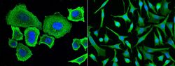
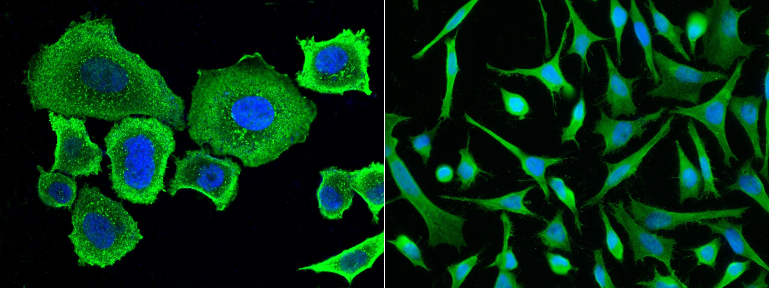
- Immunofluorescence staining of Human Angiotensinogen in Hela or SKBR3 cells. Cells were fixed with 4% PFA, permeabilzed with 1% Triton X-100 in PBS, blocked with 10% serum, and incubated with Angiotensinogen Recombinant Rabbit Monoclonal Antibody (1) (Product # MA5-29009, 1:60). Then cells were stained with the Alexa Fluor® 488-conjugated Goat Anti-rabbit IgG secondary antibody (left panel, captured by laser confocal scanning microscope; right panel, captured by fluorescence microscope), counterstained with DAPI (blue). Positive staining was localized to cytoplasm.
- Submitted by
- Invitrogen Antibodies (provider)
- Main image

- Experimental details
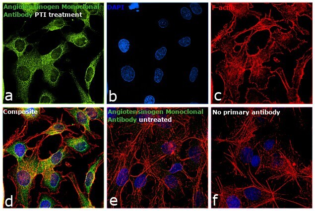
- Immunofluorescence analysis of Angiotensinogen was performed using 70% confluent log phase Hep G2 cells treated with 1X protein transport inhibitor (Product # 00-4980-03) for 4 hours. Angiotensinogen is readily secreted out in the medium after translation and post processing. Treatment of cells with protein transport inhibitor sequesters Angiotensinogen in the endoplasmic reticulum. The cells were fixed with 4% paraformaldehyde for 10 minutes, permeabilized with 0.1% Triton™ X-100 for 15 minutes and blocked with 2% BSA for 1 hour at room temperature. cells were labeled with Angiotensinogen Monoclonal Antibody (Product # MA5-29009) at 1:200 dilution in 0.1% BSA, incubated at 4 degree Celsius overnight and then labeled with Goat anti-Rabbit IgG (H+L), Superclonal™ Recombinant Secondary Antibody, Alexa Fluor 488 (Product # A27034) at a dilution of 1:2000 for 45 minutes at room temperature (Panel a: green). Nuclei (Panel b: blue) were stained with ProLong™ Diamond Antifade Mountant with DAPI (Product # P36962). F-actin (Panel c: red) was stained with Rhodamine Phalloidin (Product # R415). Panel d represents the merged image showing ER membrane/cytoplasmic localization. Panel e represents untreated Hep G2 cells. Panel f represents control cells with no primary antibody to assess background. The images were captured at 60X magnification.
- Submitted by
- Invitrogen Antibodies (provider)
- Main image

- Experimental details
- Immunofluorescence staining of Human Angiotensinogen in Hela or SKBR3 cells. Cells were fixed with 4% PFA, permeabilzed with 1% Triton X-100 in PBS, blocked with 10% serum, and incubated with Angiotensinogen Recombinant Rabbit Monoclonal Antibody (1) (Product # MA5-29009, 1:60). Then cells were stained with the Alexa Fluor® 488-conjugated Goat Anti-rabbit IgG secondary antibody (left panel, captured by laser confocal scanning microscope; right panel, captured by fluorescence microscope), counterstained with DAPI (blue). Positive staining was localized to cytoplasm.

- Submitted by
- Invitrogen Antibodies (provider)
- Main image

- Experimental details
- Immunofluorescence analysis of Angiotensinogen was performed using 70% confluent log phase Hep G2 cells treated with 1X protein transport inhibitor (Product # 00-4980-03) for 4 hours. Angiotensinogen is readily secreted out in the medium after translation and post processing. Treatment of cells with protein transport inhibitor sequesters Angiotensinogen in the endoplasmic reticulum. The cells were fixed with 4% paraformaldehyde for 10 minutes, permeabilized with 0.1% Triton™ X-100 for 15 minutes and blocked with 2% BSA for 1 hour at room temperature. cells were labeled with Angiotensinogen Monoclonal Antibody (Product # MA5-29009) at 1:200 dilution in 0.1% BSA, incubated at 4 degree Celsius overnight and then labeled with Goat anti-Rabbit IgG (Heavy Chain), Superclonal™ Recombinant Secondary Antibody, Alexa Fluor 488 (Product # A27034) at a dilution of 1:2000 for 45 minutes at room temperature (Panel a: green). Nuclei (Panel b: blue) were stained with ProLong™ Diamond Antifade Mountant with DAPI (Product # P36962). F-actin (Panel c: red) was stained with Rhodamine Phalloidin (Product # R415). Panel d represents the merged image showing ER membrane/cytoplasmic localization. Panel e represents untreated Hep G2 cells. Panel f represents control cells with no primary antibody to assess background. The images were captured at 60X magnification.

Supportive validation
- Submitted by
- Invitrogen Antibodies (provider)
- Main image
- Experimental details
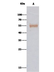
- Angiotensinogen Immunoprecipitation using: Lane A: 0.5 mg HepG2 Whole Cell Lysate 2 µL with Angiotensinogen Recombinant Rabbit Monoclonal Antibody (1) (Product # MA5-29009) and 15 µL of 50 % Protein G agarose. Primary antibody: Angiotensinogen Recombinant Rabbit Monoclonal Antibody (1), at 1:200 dilution. Secondary antibody: Clean-Bloto IP Detection Reagent (HRP) at 1:500 dilution. Developed using the DAB staining technique. Performed under reducing conditions. Predicted band size: 51 kDa. Observed band size: 51 kDa.
Supportive validation
- Submitted by
- Invitrogen Antibodies (provider)
- Main image

- Experimental details
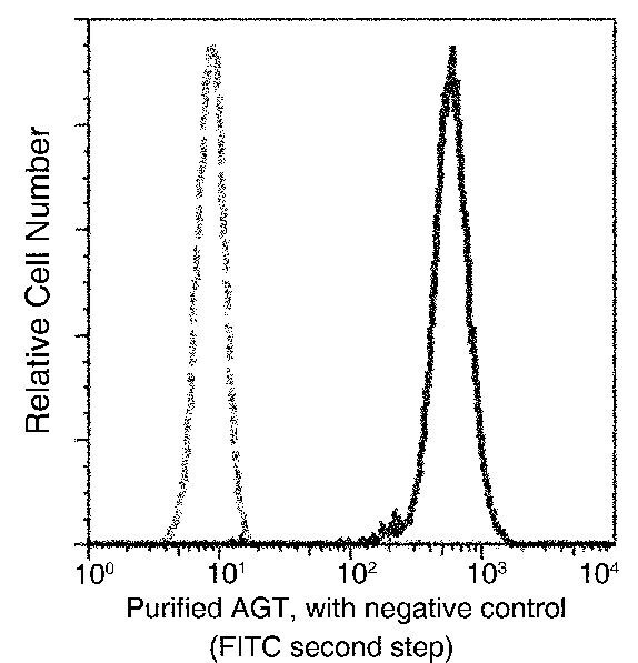
- Flow cytometric analysis of Human Angiotensinogen expression on HepG2 cells. The cells were treated according to manufacturer’s manual, stained with Angiotensinogen Recombinant Rabbit Monoclonal Antibody (1) (Product # MA5-29009), then a FITC-conjugated Secondary antibody. The fluorescence histograms were derived from gated events with the forward and side light-scatter characteristics of intact cells.

- Submitted by
- Invitrogen Antibodies (provider)
- Main image

- Experimental details
- Flow cytometric analysis of Human Angiotensinogen expression on HepG2 cells. The cells were treated according to manufacturer’s manual, stained with Angiotensinogen Recombinant Rabbit Monoclonal Antibody (1) (Product # MA5-29009), then a FITC-conjugated Secondary antibody. The fluorescence histograms were derived from gated events with the forward and side light-scatter characteristics of intact cells.
Supportive validation
- Submitted by
- Invitrogen Antibodies (provider)
- Main image

- Experimental details
- Angiotensinogen Immunoprecipitation using: Lane A: 0.5 mg HepG2 Whole Cell Lysate 2 µL with Angiotensinogen Recombinant Rabbit Monoclonal Antibody (1) (Product # MA5-29009) and 15 µL of 50 % Protein G agarose. Primary antibody: Angiotensinogen Recombinant Rabbit Monoclonal Antibody (1), at 1:200 dilution. Secondary antibody: Clean-Bloto IP Detection Reagent (HRP) at 1:500 dilution. Developed using the DAB staining technique. Performed under reducing conditions. Predicted band size: 51 kDa. Observed band size: 51 kDa.
